Download
1 / 21
270 likes | 1.03k Vues
Lecture 16: Regulation of Proteins 3: Isozymes and Covalent Modification. Isozymes Covalent Modification Protein Kinase A. Biological Processes are Carefully Regulated. Allosteric Control: The activity of some proteins can be controlled by modulating

E N D
Lecture 16:Regulation of Proteins 3:Isozymes and Covalent Modification Isozymes Covalent Modification Protein Kinase A
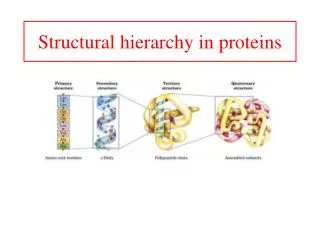
Biological Processes are Carefully Regulated Allosteric Control: The activity of some proteins can be controlled by modulating the levels of small signalling molecules. The binding of these molecules causes conformational changes in the protein which affect its activity. Multiple forms of Enzymes: Different tissues or developmental stages sometimes have specific versions of a given enzyme which have distinct properties although they may have the same basic activity. Reversible Covalent Modification: The activity of many proteins is controlled by attachment of small chemical groups. The most common such modification is phosphorylation- attachment of a phosphate group. Proteolytic Activation: Some enzymes are synthesized in an inactive form and must be activated by cleavage of the inactive form.
Multiple Forms of Proteins Different tissues or developmental stages differ in their requirements for the activity of various proteins. An example is provided by fetal hemoglobin. Oxygen is provided to the fetus through the mother’s circulatory system, which requires that oxygen be transferred from the maternal hemoglobin to the hemoglobin in the bloodstream of the fetus. The fetus expresses a different form of hemoglobin which has a higher oxygen affinity than adult hemoglobin, facilitating this transfer. This regulatory strategy is also a common means of controlling the activity of enzymes. Enzymes that carry out the same chemical reaction, but differ in sequence, kinetic properties, and/or in regulatory properties, are called isozymes.
Isozymes of Lactate Dehydrogenase Lactate dehydrogenase (LDH) functions in glucose metabolism. Mammals have 2 versions of LDH, the H isozyme (found in heart) and the M isozyme (found in skeletal muscle) which are closely related, sharing about 75% sequence identity. LDH functions as a tetramer, but individual tetramers can be composed of any combination of M and H isozymes. (M4, M3H, M2H2, MH3, or H4) The M4 tetramer functions optimally under anaerobic conditions, while the H4 tetramer functions optimally under aerobic conditions. The H4 tetramer has higher substrate affinity and also can be allosterically inhibited by pyruvate. The M4 tetramer has a lower affinity for substrate and is not allosterically regulated. The mixed tetramers exhibit properties intermediate between the the H4 and M4 tetramers- differential expression of the two isozymes allows fine control over the LDH activity.
H4 M4 Age (days) Developmental Regulation of LDH Isozymes At different developmental stages in the rat heart, the two isozymes of LDH are expressed to different degrees. In the early embryo the M form predominates, and in the adult the H form predominates. (anaerobic) (aerobic)
Tissue-Specific LDH Isozyme Expression The levels of the different isozymes also vary from tissue to tissue. There is a distribution of different complexes in a given tissue. LDH-1 LDH-2 LDH-3 LDH-4 LDH-5 Disruption of cells can lead to release of LDH into the bloodstream. In blood, the level of LDH-2 is normally higher than the level of LDH-1. But during a heart attack, LDH-1 can be released into the bloodstream, increasing it above the level of LDH-2. Analysis of the relative levels of LDH isozymes serves as a diagnostic for a variety of medical conditions.
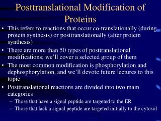
Covalent Modification The activity of many proteins is controlled by various forms of covalent modification- attachment of a small functional group. The modification can increase or suppress the activity of the proteins. Specific enzymes carry out these modifications, and other enzymes remove them. In most cases the modification is reversible- removal of the attached group reverses the effect of the modification. In this way the regulated proteins can be “turned on” and “turned off” as appropriate, depending on the cell’s need for that particular activity.
Examples of Covalent Modification A variety of modifications exist, acting to control a wide range of cellular functions.
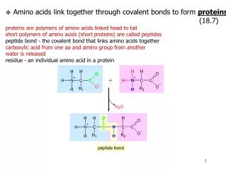
Acetylation of Histones Histones are important eukaryotic proteins involved in the packaging of DNA in chromosomes and also in regulation of gene expression. Histones DNA Section of chromatin fiber Histones are basic proteins, rich in Lys and Arg residues. The positive charge assists in binding DNA, which is negatively charged. The lysine residues of histones near regions of actively transcribed genes are heavily acetylated.
Acetylation of lysines in histones removes the positive charge, weakening the affinity of histones for DNA. This makes it easier to remove or displace the histone from DNA to enable genes to be transcribed. The covalent modfication of histones is carried out by enzymes called histone acetyltransferases. Removal of the acetyl group is carried out by deacetylases. These enzymes allow changes in the expression level of genes in various locations on the chromosome. In turn, the acetyltransferases and deacetylases are themselves regulated by phosphorylation.
Phosphorylation Probably the most widespread method of regulation is phosphorylation- attachment of a phosphate group. A wide variety of enzymes, many membrane channels and transcription factors, and virtually all metabolic processes are regulated by phosphorylation. Phosphorylation is carried out by protein kinases, enzymes which transfer a phosphate group from ATP to hydroxyl groups in proteins.
Kinases A kinase is any enzyme which transfers a phosphate group. Kinases are named for the target which receives the phosphate. (eg glycerol kinase phosphorylates glycerol) The phosphate group derives from ATP, which is only present inside cells. Extracellular proteins are not regulated by phosphorylation. An enzyme that phosphorylates a protein is a protein kinase. Two major classes of protein kinases: Serine/threonine kinases Group that receives the phosphate is a serine or threonine hydroxyl. Tyrosine kinases. Group that receives the phosphate is a tyrosine hydroxyl.
Examples of Protein Kinases Protein kinases comprise one of the largest protein families known- there are more than 500 in humans. These allow regulation specific to particular tissues, developmental stages, and substrate proteins. Kinases themselves are controlled by many different kinds of cellular signals including by other kinases.
Kinases and Phosphatases Phosphorylation is carried out by kinases. Removal of phosphate groups is carried out by phosphatases. These reactions are not the reverse of one another. Both reactions are energetically downhill and so are unidirectional but proceed extremely slowly in the absence of enzymes. Transfer of phosphate from ATP to protein. Kinase Transfer of phosphate from protein to water. Phosphatase
Effects of Phosphorylation Phosphorylation often activates a target molecule, for example by inducing a conformational change to a more active state. The influence on target activity can be accomplished through various effects. Charge: Phosphorylation adds 2 negative charges which can participate in (or disrupt) charge-charge interactions. Hydrogen bonding: The phosphate group can participate in 3 or more hydrogen bonds. Energy: The substantial amount of energy in the phosphate bond can strongly affect conformational equilibria. Time: Phosphorylation can be accomplished in seconds and can last for as long as needed. By regulating the phosphorylation and dephosphorylation steps, the activity of the target can be adjusted to synchronize with a physiological process. Amplification: A single kinase can phosphorylate and activate hundreds of target molecules resulting in a large effect from a small stimulus.
Specificity of Protein Kinases Dedicated protein kinases phosphorylate only a single target or a few closely related ones, allowing fine control over this limited target. Multifunctional protein kinases phosphorylate many different targets, allowing a single kinase to control a variety of different processes. For kinases with many different targets, comparison of the amino acid sequences of the residues near the phosphorylation sites identifies patterns. The primary determinant of specificity is the amino acids surrounding the phosphorylation site. For example, protein kinase A recognizes and phosphorylates the serine or threonine in the sequences: Arg-Arg-X-Ser-Z or Arg-Arg-X-Thr-Z where X is a small residue (eg Gly) and Z is a large hydrophobic residue (eg Ile)
Protein Kinase A Many physiological processes are regulated by hormones, which are are extracellular signalling molecules. Hormones bind to receptors at the cell surface. In some cases, the binding of hormones causes the formation of intracellular signalling molecules, or second messengers, such as cyclic AMP. This carries the signal from the hormone into the cell and results in activation of many different proteins. Most of these proteins are activated by a single kinase, protein kinase A. (PKA)
Protein Kinase A is Regulated by cAMP PKA consists of: 2 C subunits (catalytic subunits) that contain the kinase activity 2 R subunits (regulatory subunits) that bind cAMP In the absence of cAMP, an inactive C2R2 complex is formed in which the regulatory subunits tightly bind and sequester the catalytic subunits. In the presence of cAMP (eg in response to a hormone) the regulatory subunit binds the cAMP, inducing a conformational change that releases the catalytic subunits, which can then begin phosphorylating their targets.
Mechanism of cAMP Regulation The R subunit of PKA contains a sequence nearly matching the preferred substrate of the C subunits but incapable of being phosphorylated. This pseudo-substrate sequence contains the residues …Arg-Arg-Gly-Ala-Ile… and the C subunit binds this sequence tightly at its active site, preventing other substrates from being phosphorylated. The binding of cAMP by the R subunit causes an allosteric conformational shift which removes the pseudo-substrate sequence from the C subunit, releasing it.
Structural Basis of cAMP Regulation The structure of the catalytic subunit of PKA in complex with a pseudosubstrate peptide allows the substrate specificity and inhibition by the R subunit to be understood as due to complementarity with specific residues in the c subunit. Arg-Arg-Asn-Ala-Ile Ionic interactions Hydrophobic interactions Catalytic subunit
Summary: Isozymes are enzymes which have the same activity but different kinetics or regulatory properties- differential expression of isozymes allows control over enzyme activity. Many proteins are regulated by covalent modification. The most common such modification is phosphorylation. Protein kinase A carries out phosphorylation of a wide variety of targets in response to cyclic AMP. Key Concepts: Isozymes of LDH Regulation of histones by acetylation Kinases Phosphatases Role of Protein Kinase A