Download
1 / 48
490 likes | 944 Vues
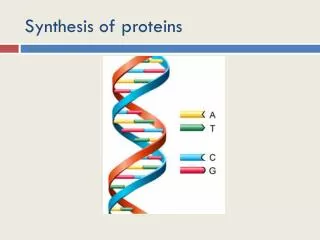
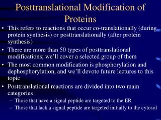
Posttranslational Modification of Proteins. This refers to reactions that occur co-translationally (during protein synthesis) or posttranslationally (after protein synthesis) There are more than 50 types of posttranslational modifications; we’ll cover a selected group of them

E N D
Posttranslational Modification of Proteins • This refers to reactions that occur co-translationally (during protein synthesis) or posttranslationally (after protein synthesis) • There are more than 50 types of posttranslational modifications; we’ll cover a selected group of them • The most common modification is phosphorylation and dephosphorylation, and we’ll devote future lectures to this topic • Posttranslational reactions are divided into two main categories • Those that have a signal peptide are targeted to the ER • Those that lack a signal peptide are targeted initially to the cytosol
Protein Targeting Nascent polypeptide/ribosome - Signal Seq + Endoplasmic Reticulum Cytosol Plasma Membrane Golgi Secretory Vesicles Mitochondria Nucleus Lysosomes Plasma Membrane Cytosolic Pathway Secretory Pathway No signal peptide With a signal peptide
Cytosolic Pathway • Proteins that lack a signal peptide at the amino terminus are not translocated into the Golgi and are not processed • These proteins are synthesized on free ribosomes not associated with the rough endoplasmic reticulum • Final cell location • Cytosol, e.g., hexokinase • Nucleus, e.g., DNA polymerase • Mitochondrion, e.g., cytochrome c • Other modifications in the cytosol • Acetylation (2C) Prenylation (15 or 20C) • Myristoylation (14 C) Palmitoylation (16C)
NLS (Nuclear Localization Sequence) • The nucleus is surrounded by a nuclear envelope • Inner nuclear membrane • Outer nuclear membrane • Macromolecules are translocated through a nuclear pore • All proteins found in the nucleus are synthesized in the cytosol and are translocated through the nuclear pore into the nucleus • Histones, DNA polymerases, RNA polymerases • Transcription factors, splicing factors
NLS (Nuclear Localization Sequence) • Nuclear proteins contain an NLS • One or two sequences (patches) rich in lysine and arginine • Can be found anywhere in the protein; at the N-terminus, in the middle, or at the C-terminus • PKKKRKV is an example; PKNKRKV is inactive • Attachment of this sequence to normally cytosolic proteins results in the import of such mutated proteins into the nucleus • The nucleoplasminin story • It is required for chromatin assembly • It contains two patches that are required for nuclear import • A Lys-Arg pair • Four lysines located 10 amino acids further downstream • KRPAATKKAGQAKKKK, where the key residues are underlined
NLS (Nuclear Localization Sequence) • Mechanism • Proteins with an NLS bind to importins that take them to the nuclear pore • The alpha subunit of importin binds the NLS • The beta subunit binds to the nuclear pore • A Ran GTPase interacts with the protein-importin complex and energizes nuclear translocation with GTP hydrolysis • The NLS is not removed proteolytically. Why? • Nuclear Export Sequence (NES) • Newly synthesized ribosomes (RNA and protein) bear nuclear export sequences • This may involve signals on both proteins and RNA • Nuclear Retention Signal (NRS) • Found in proteins that bind to immature RNAs in the nucleus • mRNAs that contain introns • Pre-tRNAs
Nuclear Import and Export • Importin binds to cargo and interacts with nucleoporins • It is a nuclear import receptor • It binds to basic NLSs • RanGTP occurs in the nucleus, RanGDP in the cytosol • RanGTP dissociates import complexes • RanGTP forms export complexes • Ran guanine nucleotide exchange factor (RanGEF) occurs in the nucleus • RanGTPase activating protein (RanGAP) occurs in the cytosol • Important points • Ran is a GTPase • RanGTP is nuclear • RanGDP is cytosolic
Mitochondria and Protein Import • Powerhouse of the cell • Krebs cycle, beta oxidation, pyruvate dehydrogenase • Contain DNA, RNA, ribosomes • Synthesize about 20 mitochondrial proteins • Most mitochondrial proteins are synthesized in the cytosol and imported
Import of Proteins • Amino-terminal sequence (10-70 aa) contains positively charged, ser/thr, and hydrophobic amino acids but no common sequence • Cyt c has an internal targeting sequence • Hsp70 keeps proteins in an unfolded state • Translocation through TOM (transport outer membrane) • The fit is snug during transport • Ions and other small molecules do not leak across the membrane • Voltage gradient is required for transport across the inner membrane by TIM • Presequence is cleaved by a signal protease • Mit Hsp 70 and 60 aid in translocation and facilitates folding
Insertion of mit membrane proteins • Proteins targeted for mitochondrial membranes contain hydrophobic stop sequences that halt translocation through the TOM or TIM complexes
Sorting proteins to the intermembrane space • I Through Tom into inner mitochondrial space • II From Tom to Tim with hydrophobic stop sequences that are cleaved • III Into matrix • Remove hydrophilic basic sequence • Exposes hydrophobic sequence that directs protein to inner mitochondrial space
Protein Import and the Peroxisome • Peroxisomes oxidize lipids (fatty acids > 18 carbon atoms) • Peroxisomes contain a single lipid bilayer membrane • Unlike mitochondria, peroxisomes lack DNA • All proteins are encoded by nuclear genes • Peroxisome Targeting Signal (PTS) • Type I; PTS1 • C-terminus • Ser-Lys-Ala (Don’t memorize) • Type II; PTS2 • Rare (4 in humans) • At or near the N-terminus • RLXXXXXH/QL (Don’t memorize) • Peroxins deliver peroxisomal proteins to the target and insert them into the matrix or the membrane (mechanism ?) • Take home message: there are peroxisome targeting signals
Membrane Localization Signals I • Posttranslational attachment of lipids to proteins creating non-membrane spanning integral membrane proteins that will reside on the cytoplasmic surface of the plasma membrane of subcellular membranous organelle • Myristoylation (14C) • N-terminal processing • Met-aminopeptidase often removes N-terminal Met • If residue after methionine is a glycine, a myristoyl group can be attached via an amide linkage, blocking the amino terminal group • The lipophilic myristoyl group can be inserted into the membrane • Several of the alpha subunits of heterotrimeric G-proteins possess this modification • Not all proteins that are N-myristoylated are attached to membranes • Myristoyl~CoA + H2N-Gly-protein myristoyl-CO-N(H)-Gly-protein + CoA; the high energy thioester is used to drive the synthesis of the low energy amide linkage
Protein Prenylation II • A 15 carbon farnesyl group or a 20 carbon geranylgeranyl group is added to proteins that contain a C-terminal CaaX box • C is cysteine • a represents aliphatic residues (not Alanine) • X represents leucine for geranylgeranyl groups and Met, Ser, Ala for farnesylation • Ras is farnesylated • Part of the Raf-MEK-ERK pathway • Mutated in 25% of all human cancers • Inhibition of Ras farnesylation is a targeted anticancer target • The gamma subunit of many G-gamma proteins is geranylgeranylated • These modifications promote membrane binding • Know what a CaaX box is
Prenylation Sequence of Reactions Fig. 18-17
Palmitoylation Reactions (16C) • K-ras, one type of ras, is both farnesylated and palmitoylated • A protein cysteine is modified as a thioester • Palmitoyl-CoA + protein CysSH protein CysS~palmitate + CoA • This concludes the Cytosolic Pathway • Next, the Secretory pathway
Secretory Pathway • Products for secretion, transmembrane proteins, and import into Golgi/ER/Secretory granules • Preproinsulin (secreted) • Prealbumin (secreted) • Preproinsulin receptor beta subunit (transmembrane) • The pre refers to the signal peptide • A signal peptide pre sequence at the amino terminus of a protein targets polypeptide/ribosome to the ER • 6-13 hydrophobic proteins near the N-terminus • Usually a positively charged residue nearby • This is the second sequence besides CaaX that you should learn • Some proteins are cleaved by signal peptidases and are found entirely within the lumen • Some proteins contain “stop transfer” sequences • These proteins become membrane spanning portions of integral membrane proteins
Protein Synthesis • We’ll see this again when we cover amelogenin biosynthesis in enamel formation • Fig. 18-4
Role of the Signal Sequence and Signal Recognition Particle in Directing a Peptide to the ER (Fig. 18-2) Fig. 18-2
Insulin Biosynthesis • Synthesized as preproinsulin, the first such discovered protein • The presequence is cleaved to yield proinsulin (signal peptidase) • Disulfide bonds form, and the connecting peptide is cleaved by prohormone convertase • Carboxypeptidase H finishes the job by cleaving the basic residues • This yields mature insulin with its A and B chains • Learn this process; it’s an important prototype • Fig. 18-3
GPI Anchor Biosynthesis • GPI: GlycoPhosphatidyl-Inositol anchor • Fatty acids in membrane • Polar groups in lumen • GPI transamidase catalyzes the reaction of the amino group with a protein carboxylate to give a new amide bond • …C(=O)-N(H)-.. + H2N- …C(=O)-N(H)-.. + H2N-
Cotranslational and Posttranslational Modifications in the ER and Golgi • Most of the modifications produced in the ER are constitutive (remain until the protein is degraded) • These modifications take advantage of the unfolded nature of the polypeptide as it enters the lumen of the ER • Glycosylation Reactions attach carbohydrate: O-linked and N-linked (Fig. 18-7) • O-linked refers refers to the attachment of sugars to serine or threonine (simple) • N-linked refers to the attachment of sugars to asparagine (difficult) • High mannose • Complex • Hybrid
Endoplasmic Reticulum and Golgi • Endoplasmic (inside the cell); reticulum a network • ER, a network inside the cell • Disulfide bond formation occurs in the ER • N-linked oligosaccharide synthesis is initiated in the ER; trimming and completion occurs in the Golgi • Most O-glycosylation occurs in the Golgi • Attachment of mannose 6-phosphate occurs in the Golgi • Sulfation of secreted proteins occurs in the Golgi • Proline and lysine hydroxylation, alpha amidation, and vitamin K-dependent carboxylation reactions occur in the Golgi
Activated Carbohydrates • These serve as carbohydrate donors • As activated sugars, a high-energy bond is used for the synthesis of a low-energy compound • UDP-Glu, UDP-Gal, UDP-GlcUA, UDP-Xyl, UDP-GlcNAc, UPD-GalNAc, GDP-Man • CMP-NeuNAc • Dolichol phosphates • It is not necessary to memorize the following pathways, but you should remember the identity of the activated sugars
First Stage of N-Linked Oligosaccharide Synthetsis (Fig. 18-12): Occurs in the ER
Second and Third Stages of N-Linked Oligosaccharide Synthesis (Fig. 18-13) • The hydrolysis reactions are unidirectional • Donation of activated sugars is energetically favorable • Each reaction is catalyzed by an enzyme that determines the sequence of sugars and the configuration of the glycosidic linkages • Occurs in Golgi
Blood Group Biosynthesis • Fig. 18-16 • Golgi reactions • People with type O blood groups lack functional A and B genes • Genes A and B differ by 4 nucleotides which alters the substrate specificity A: GalNAc transferase B: Gal transferase
Targeting Enzymes to Lysosomes:A Golgi Process • Lysosomal proteins contain N-linked oligosaccharides with terminal mannose 6-phosphates • The addition of phosphate occurs by an unusual mechanism or pathway • There is a mannose 6-phosphate receptor that recycles between the Golgi and lysosome and participates in the translocation of lysosomal enzymes
Protein Sulfation Reactions (Fig. 12-12) Active sulfate: PAPS, phosphoadenosylphosphosulfate
Protein Sulfation • A Golgi pathway modification • Fig. 18-6
Procollagen Hydroxylation • Fig. 18-18 • Golgi reactions • Proline and lysine hydroxylation reactions • Requires vitamin C • These two hydroxylases • Dopamine beta-hydroxylase • Peptidyl amidating mono-oxygenase • The lysyl oxidase Rxn (next slide) inititates collagen cross linking
Lysyl Oxidase Reaction (Fig. 23-12) Golgi reactions
Protein Amidation • The amide group is derived from a carboxyterminal glycine • Ascorbate and oxygen are required • Golgi reactions • Fig. 18-19
Vitamin K-Dependent Carboxylation Reactions • Protein-glutamate is carboxylated • Carbon dioxide, Vitamin K, and oxygen are required • Several blood clotting factors and other proteins that bind calcium ions contain gamma carboxylation of protein-glutamates • Golgi reactions
Plasma Membrane Topology • It is not necessary to remember which protein has which type of membrane topology except for GPCRs
Topological Classes • Topological classes I-III have a single pass through the membrane • I: N-terminus is intraluminal and C-terminus is extraluminal • II: C-terminus is extra, N-terminus is intra; no cleaved sequence • III: Same as I, but no cleavable sequence • Class IV has multiple passes • Type I, without a signal peptide, contains a 22 aa hydrophobic stop transfer sequence • Type II and III • Lack a N-terminal signal peptide • Contain a signal-anchor sequence that functions as an ER signal sequence and membrane anchor sequence
Type II Membrane Protein Biosynthesis • Stop transfer anchor sequence • C-terminus in the ER lumen or cell exterior
Protein Targeting Nascent polypeptide/ribosome - Signal Seq + Endoplasmic Reticulum Cytosol Plasma Membrane Lipidation with myristate, palmitate, farnesylate, or geranylgeranylate Golgi Glycosylation, Amidation, Sulfation, K carbox, Hydroxylation Mitochondria Basic aa at N-ter Nucleus Basic amino acids Secretory Vesicles Lysosomes Mannose 6-P Plasma Membrane Stop transfer sequences