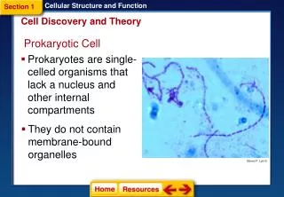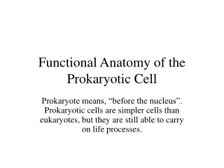
Prokaryotic Cell
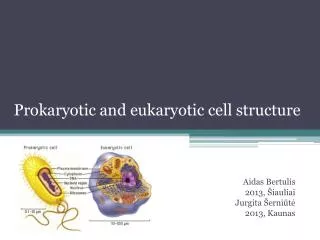
Prokaryotic Cell . Eukaryotic Cell. “true” nucleus Membrane bound organelles 1.5 billion years Both unicellular and multicellular Linear DNA Some have cell walls, some don't. “before” Nucleus (has no nucleus) No membrane bound organelles 3.5 billion years Unicellular Circular DNA

Prokaryotic Cell
E N D
Presentation Transcript
Prokaryotic Cell Eukaryotic Cell “true” nucleus Membrane bound organelles 1.5 billion years Both unicellular and multicellular Linear DNA Some have cell walls, some don't • “before” Nucleus (has no nucleus) • No membrane bound organelles • 3.5 billion years • Unicellular • Circular DNA • Contain a cell wall
Prokaryotic Cell Eukaryotic Cell “true” nucleus Membrane bound organelles 1.5 billion years Both unicellular and multicellular Linear DNA Some have cell walls, some don't • “before” Nucleus (has no nucleus) • No membrane bound organelles • 3.5 billion years • Unicellular • Circular DNA • Contain a cell wall
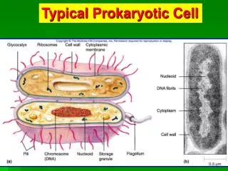
Characteristics • Prokaryotes • Microscopic (Eukaryotic cells are at least 10x bigger) • Unicellular • DNA is a single circular piece of DNA • Asexual Reproduction • Binary Fission • Genetic Exchange • Conjugation –transfer DNA through contact • Transformation – acquire DNA from dead bacteria • Transduction – DNA is transferred from one bacteria to another using a virus (genetic engineering) • Metabolism • Aerobic • Anaerobic
http://highered.mcgraw-hill.com/sites/0072556781/student_view0/chapter13/animation_quiz_3.htmlhttp://highered.mcgraw-hill.com/sites/0072556781/student_view0/chapter13/animation_quiz_3.html http://highered.mcgraw-hill.com/sites/0072556781/student_view0/chapter13/animation_quiz_2.html
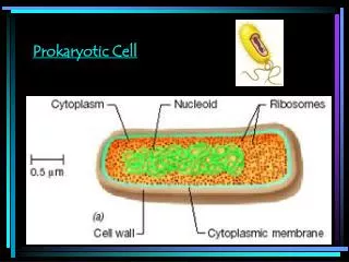
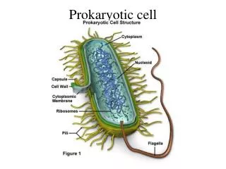
Structure • Pilus (Pili)- allows them to adhere to surfaces • Flagellum – movement • Cell Wall – Made of peptidoglycan; Used in medicine to identify type of bacterium using Gram Stain (pg. 463)
Gram Stain (pg. 529) • Gram + simple walls, large amount of peptidoglycan • Gram - less peptidoglycan, outer membrane contains lipopolysaccharides which are often toxic and provides additional protection more resistant to antibiotics • Many antibiotics (penicillens) inhibit synthesis of cross links in peptidoglycan and prevent formation of a functional wall Gram negative Gram positive
Gram Positive Organisms • Aerobic, Gram-positive cocci • Staphylococcus aureus (fig 1, 2, 3, 4) • Staphylococcus epidermidis (fig 1) • Staphylococcus sp. (Coagulase-negative)(fig 1) • Streptococcus pneumoniae (Viridans group)(fig 1, 2, 3) • Streptococcus agalactiae (group B)(fig 1) • Streptococcus pyogenes (group A)(fig 1, 2) • Enterococcus sp.(fig 1, 2, 3) • Aerobic, Gram-positive rods • Bacillus anthracis (fig 1, 2) • Bacillus cereus (fig 1, 2) • Bifidobacterium bifidum (fig 1) • Lactobacillus sp. (fig 1, 2) • Listeria monocytogenes (fig 1, 2) • Nocardia sp.(fig 1, 2) • Rhodococcus equi (coccobacillus)(fig 1) • Erysipelothrix rhusiopathiae (fig 1) • Corynebacterium diptheriae (fig 1, 2) • Propionibacterium acnes (fig 1) • Anaerobic, Gram-positive rods • Actinomyces sp. (fig 1, 2) • Clostridium botulinum (fig 1) • Clostridium difficile (fig 1) • Clostridium perfringens (fig 1, 2, 3) • Clostridium tetani (fig 1, 2) • Anaerobic, Gram-positive cocci • Peptostreptococcus sp. (fig 1)
Gram Negative Organisms • Aerobic, Gram-negative cocci • Neisseria gonorrhoeae (fig 1, 2, 3, 4) • Neisseria meningitidis (fig 1; false color of the bacterium., 2) • Moraxella catarrhalis (fig 1) • Anaerobic, Gram-negative cocci • Veillonella sp. (fig 1) • Aerobic, Gram-negative rods • Fastidious, Gram-negative rods • Actinobacillus actinomycetemcomitans (fig 1) • Acinetobacter baumannii(fig 1 really A. calcoaceticus) • Bordetella pertussis (fig 1, 2) • Brucella sp. (fig 1) • Campylobacter sp.(fig 1) • Capnocytophaga sp.(fig 1,2) • Cardiobacterium hominis (fig 1) • Eikenella corrodens (fig 1) • Francisella tularensis (fig 1,) • Haemophilus ducreyi (fig1,2) • Haemophilus influenzae (fig 1, 2) • Helicobacter pylori (fig 1, 2, 3, 4) • Kingella kingae (fig ) • Legionella pneumophila (fig 1, 2, 3) • Pasteurella multocida (fig 1) • Enterobacteriaceae (glucose-fermenting Gram-negative rods) • Citrobacter sp. (fig 1) • Enterobacter sp. (fig 1) • Escherichia coli (fig 1, 2) • Klebsiella pneumoniae (fig1, 2) • Proteus sp. (fig 1) • Salmonella enteriditis (fig 1) • Salmonella typhi (fig 1) • Serratia marcescens (fig 1, 2) • Shigella sp. (fig 1) • Yersinia enterocolitica (fig 1) • Yersinia pestis (fig 1, 2) • Oxidase-positive, glucose-fermenting Gram-negative rods • Aeromonas sp. (fig 1) • Plesiomonas shigelloides (fig 1) • Vibrio cholerae (fig 1, 2) • Vibrio parahaemolyticus (fig 1) • Vibrio vulnificus (fig 1) • Glucose-nonfermenting, Gram-negative rods • Acinetobacter sp. (fig 1) • Flavobacterium sp. (fig 1) • Pseudomonas aeruginosa (fig 1, 2) • Burkholderia cepacia (fig 1) • Burkholderia pseudomallei (fig 1) • Xanthomonas maltophilia or Stenotrophomonas maltophila(fig 1) • Anaerobic, Gram-negative rods • Bacteroides fragilis (fig 1) • Bacteroides sp. (fig 1) • Prevotella sp. (fig 1) • Fusobacterium sp. (fig 1,2) • Gram-negative spiral • Spirillum minus (minor)- (fig 1)
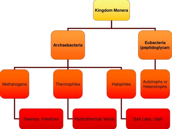
Nutrition • Autotrophic • Photosynthetic • Chemoautotrophic (nitrogen fixers) • Heterotrophic • Decomposer • Parasitic (Treponema pallidum)
Survival of the Fittest!!! • Bacteria have been around for 3.5 billion years!! How???? • Cell Walls • Capsules (surrounds cell wall) • Endospores : allow them to withstand drought, high temps., lack of food, etc. • Super fast reproduction • Asexual Reproduction, but can still acquire other genes • Inhabit every place on Earth
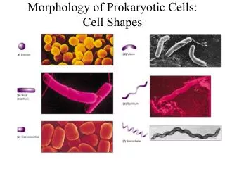
Classification • Arrangements • Strept : Chains • Staph : Clusters • Diplo : Pairs • Shapes • Coccus : Spheres • Bacillus : Rods • Spirillum : Spirals
Importance to Humans??? • Bacteria are used to make food • Pickles, buttermilk, cheese, sauerkraut, olives, vinegar, sourdough bread, beer, wine • Bacteria can produce chemicals • Acetone, Butanol • Important Recyclers in environment • Nitrogen cycle • Bacteria can help clean up chemical spills • Mining companies harvest copper or uranium by using Chemoautotrophic • Bacteria are used to produce medicines • Insulin • Bacteria cause disease • Produce toxins (Clostridium botulinum) • Metabolize their host (Mycobacterium tuberculosis)
Antibiotic Resistance • Bacteria acquire genes that help the cell resist treatment using antibiotics • Arises naturally in bacteria • Occurs when weaker bacteria die off, but stronger ones survive and reproduce. • Overuse and misuse of Antibiotics has increased Ab resistance among bacteria
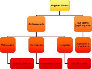
Internet Resources • Life History and Ecology of Bacteria • Bacteria • CELLS alive! Table of Contents • archaebacteria