VASCULITIS
VASCULITIS. FERDA ÖZKAN M.D. YEDITEPE UNIVERSITY MEDICAL FACULTY. OBJECTIVES. Review vessel structure Explain inflammatory processes of vessels Describe types of vasculitis.

VASCULITIS
E N D
Presentation Transcript
VASCULITIS FERDA ÖZKAN M.D. YEDITEPE UNIVERSITY MEDICAL FACULTY
OBJECTIVES • Review vessel structure • Explain inflammatory processes of vessels • Describe types of vasculitis
The basic constituents of the walls of blood vessels are endothelial cells and smooth muscle cells, and extracellular matrix (ECM), including elastin, collagen, and glycosoaminoglycans.
The three concentric layers: • -intima, • -media, • -adventitia are most clearly defined in the larger vessels, particularly arteries.
In normal arteries, the intima consists of a single layer of endothelial cells with minimal underlying subendothelial connective tissue. • It is separated from the media by a dense elastic membrane called the internal elastic lamina. • The smooth muscle cell layers of the media near the vessel lumen receive oxygen and nutrients by direct diffusion from the vessel lumen, facilitated by holes in the internal elastic membrane
However, diffusion from the lumen is inadequate for the outer portions of the media in large and medium-sized vessels, therefore these areas are nourished by small arterioles arising from outside the vessel (called vasa vasorum, literally "vessels of the vessels") coursing into the outer one half to two thirds of the media.
The outer limit of the media of most arteries is a well-defined external elastic lamina. External to the media is the adventitia, consisting of connective tissue with nerve fibers and the vasa vasorum
ARTERIES • Arteries are divided into three types: • (1) large or elastic arteries, including the aorta, • (2) medium-sized or muscular arteries, • (3) small arteries
Large or elastic arteries, including the aorta, its large branches (particularly the innominate, subclavian, common carotid, and iliac), and pulmonary arteries;
2-medium-sized or muscular arteries, comprising other branches of the aorta (e.g., coronary and renal arteries); and • 3- small arteries (less than approximately 2 mm in diameter) and arterioles (20 to 100 μm in diameter), within the substance of tissues and organs.
Capillaries, approximately the diameter of a red blood cell (7 to 8 μm), have an endothelial cell lining but no media.
VEINS • Veins have larger diameters, larger lumens, and thinner and less well organized walls • Thus, because of their poor support, veins are predisposed to irregular dilation, compression, and easy penetration by tumors and inflammatory processes.
VEINS • The venous system collectively has a large capacity; approximately two thirds of all the blood is in veins. Reverse flow is prevented by venous valves in the extremities, where blood flows against gravity.
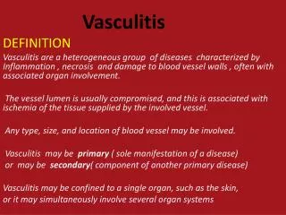
VASCULITIS • Vasculitis is a general term for vessel wall inflammation. • The clinical features of the various vasculitides are diverse and largely depend on the vascular bed affected (e.g., central nervous system vs. heart vs. small bowel).
VASCULITIS • This is a heterogenous group of disorders characterized byinflammation & damageof blood vessels followed bythrombosis & ischemic manifestationsin the tissues supplied by the blood vessels.
VASCULITIS • Besides the findings referable to the specific tissue(s) involved, the clinical manifestations typically include constitutional signs and symptoms such as fever, myalgias, arthralgias, and malaise.
INFECTIOUS VASCULITIS • Localized arteritis may be caused by the direct invasion of infectious agents, usually bacteria or fungi, and in particular Aspergillus and Mucor species.
INFECTIOUS VASCULITIS • Vascular invasion can be part of a localized tissue infection (e.g., bacterial pneumonia or adjacent to abscesses), or • less commonly-it can arise from hematogenous seeding of bacteria during septicemia or embolization from sepsis of infective endocarditis.
NONINFECTIOUS VASCULITIS • The main immunological mechanisms that initiate noninfectious vasculitis are: • (1) immune complex deposition, • (2) antineutrophil cytoplasmic antibodies, and • (3) anti-endothelial cell antibodies.
Immune Complex-Associated Vasculitis The lesions resemble those found in experimental immune complex-mediated conditions such as the Arthus reaction and serum sickness Many systemic immunological diseases, such as systemic lupus erythematosus (SLE) and polyarteritis nodosa, manifest as immune complex-mediated vasculitis.
Immune Complex-Associated Vasculitis • Antibody and complement are typically detected in vasculitic lesions, although the nature of the antigens responsible for their deposition cannot usually be determined.
Immune Complex-Associated Vasculitis • Circulating antigen-antibody complexes may also be seen (e.g., DNA-anti-DNA complexes in SLE-associated vasculitis.
Antineutrophil Cytoplasmic Antibodies Patients with vasculitis have circulating antibodies that react with neutrophil cytoplasmic antigens, so-called antineutrophil cytoplasmic antibodies (ANCAs).
Antineutrophil Cytoplasmic Antibodies ANCAs are a heterogeneous group of autoantibodies directed against constituents (mainly enzymes) of neutrophil primary granules, monocyte lysosomes, and endothelial cells
Antineutrophil Cytoplasmic Antibodies These were previously classified according to their intracellular distribution, either cytoplasmic (c-ANCA) or perinuclear (p-ANCA).
ANCAS They are discriminated based on their target antigens: Anti-myeloperoxidase (MPO-ANCA): MPO is a lysosomal granule constituent normally involved in generating oxygen free radicals. MPO-ANCAs can be induced by a variety of therapeutic agents, in particular propylthiouracil. These have been called p-ANCA.
ANCAS Anti-proteinase-3 (PR3-ANCA): PR3 is also a neutrophil azurophilic granule constituent. That it shares homology with numerous microbial peptides may explain how PR3-ANCAs develop. These have been called c-ANCA.
Mechanism for ANCA vasculitis - Drugs or cross-reactive microbial antigens induce ANCAs; alternatively, neutrophil surface expression or release of PR3 and MPO (e.g., in the setting of infections) incites ANCA formation in a susceptible host.
Mechanism for ANCA vasculitis Subsequent infection, endotoxin exposure, or other inflammatory stimuli elicit cytokines such as TNF that cause surface expression of PR3 and MPO on neutrophils and other cell types.
Mechanism for ANCA vasculitis • ANCAs react with these cytokine-activated cells and either cause direct injury (e.g., to endothelial cells) or induce further activation (e.g., in neutrophils). • ANCA-activated neutrophils degranulate and also cause injury by releasing reactive oxygen species, engendering endothelial cell toxicity and other indirect tissue injury.
Mechanism for ANCA vasculitis • ANCAs directed against constituents other than PR3 and MPO are also found in some patients with inflammatory disorders that do not involve vasculitis (e.g., inflammatory bowel disease, primary sclerosing cholangitis, rheumatoid arthritis
ANCA staining c-ANCA p-ANCA
Anti-Endothelial Cell Antibodies • Antibodies to endothelial cells may predispose to certain vasculitides, for example, Kawasaki disease
Pathology:Small vesselsHenoch-SchönleinPurpura • form of hypersensitivity vasculitis in kids, young adults • clinical: • purpura on buttocks, arms, legs • necrotizing vasculitis involving small dermal vessels • arthritis • abdominal pain – often with bloody diarrhea/other evidence of intestinal bleeding, due to mucosal/submucosal vasculitis • kidney involvement in 1/3 – proteinuria, nephrotic syndrome, gross/microscopic hematuria. Pathology: • glomerulonephritis is often focal, mesangial proliferative in type; often self-limiting • IgA is predominant Ab in glomerular and skin lesions.
Pathology:Small vesselsMixed Cryoglobulinemia syndrome • Clinical: • widespread small vessel vasculitis, often associated with severe glomerulonephritis • purpura • arthralgia or arthritis • cryoglobulins • reversibly precipitatie in the cold • consist of IgM rheumatoid factors • most seen in patients with HCV
Pathology: • vessels show deposits containing cryoglobulins and complement • leukocytoclastic vasculitis.
Pathology:Medium-sized vesselsPolyarteritisNodosa • 40-50s, May be life threatenting; fever, myalgia, weight loss, foot drop, weakness, abdominal pain, hypertension from renal arteriole involvement • mononeuritis complex - asymmetric peripheral neuropathy with sudden or subacute onset due to nerve infarction; many modalities lost in one nerve • local ischemia, inflammation of affected organs • kidney, GI tract, joints/muscles, heart, nervous system, skin, lungs may be affected.
PolyarteritisNodosa • Polyarteritis nodosa (PAN) is a systemic vasculitis of small or medium-sized muscular arteries (but not arterioles, capillaries, or venules), typically involving renal and visceral vessels but sparing the pulmonary circulation
PolyarteritisNodosa • There is no association with ANCAs, but about 30% of patients with PAN have chronic hepatitis B with HBsAg-HbsAb complexes in affected vessels, indicating an immune complex-mediated etiology
PolyarteritisNodosa • The cause remains unknown in the majority of cases; there may be etiologic and important clinical distinctions between classic idiopathic PAN, the cutaneous forms of PAN, and the PAN associated with chronic hepatitis. Clinical manifestations result from ischemia and infarction of affected tissues and organs.
Findings • Digital gangrene, ulceration • Renal - Hypertension, hematuria,renal failure • GIS - hematemesis, melena • RS - pneumonitis, pleural effusion • CNS - mononeuritis multiplexa • Skin - urticaria, palpable purpura, livedo reticularis.
Diagnosis: • microaneurysms on angiography if medium vessels are target • segmental damage to artery walls; present in up to 50% of cases • aneurysms or stenosis in mesenteric vessels in absence of atherosclerosis is very helpful to make diagnosis. • Treatment: steroids, immunosuppressive drugs, some respond to bactrim.
Pathology All stages of activity may coexist in different vessels, even in one vessel. • Early: focal, fibrinoid necrosis of artery/arteriole wall; transmural inflammation – PMN, eosinophilic poly infiltrate • Intermediate: mural/occlusive thrombi • Late: aneurysms if segmental involvement • with healing, wall infiltrated by fibroblasts -> fibrous thickening of wall -> nodular appearance.
Pathology:Large vesselsGiant cell arteritis and temporal arteritis • Mean age of onset 70 years; • Commonly associated with clinical syndrome polymyalgia rheumatica: • pain, stiffness in shoulder & pelvic girdles in absence of evidence of weakness or atrophy • ESR • response to low-moderate steroid doses. • Etiology: likely mediated by immune reactions to elastin
Granulomatous inflammatory process can affect any elastic and muscular artery: • most often seen in superficial temporal artery, other cranial arteries • chief clinical risk is blindness • clinical presentation: headache, scalp tenderness, claudication of the jaw(tired jaw on chewing), transient visual disturbances, musculoskeletal symptoms (polymyalgia rheumatica) , fever, malaise, weight loss, anemia.
Extracranial disease in 10-15%: • intermittent claudication is common • arterial bruits, blood pressure abnormalities. • Treatment: responds well to steroids. • Complications • coronary artery involvement myocardial ischemia • aortic valve incompetence • aortic dissection • aortic aneurysm, may rupture.
Pathology • Inflammation confined to media; mixed cell infiltrate – lymphocytes, macrophages • Giant cells may be present at junction of intima and media (eating internal elastic lamina) • Intimal proliferation.
Temporal (giant cell) arteritis. A, H&E stain of section of temporal artery showing giant cells at the degenerated internal elastic membrane in active arteritis (arrow )
Elastic tissue stain demonstrating focal destruction of internal elastic membrane (arrow) and intimal thickening (IT) characteristic of long-standing or healed arteritis.
Cogan's disease • Cogan's disease is another rare disease usually affecting young adults. • It features abrupt onset of • nerve deafness, • interstitial keratitis, and/or • a systemic vasculitis often with • aortic aneurysm formation. • It's apparently caused by an autoantibody against inner ear and endothelium