The Role of the Cerebrum in the Nervous System
Explore the functions and anatomy of the cerebrum, the largest part of the brain responsible for sensory and motor functions, memory, reasoning, and more. Learn about the cerebral cortex, lobes, and hemispheres that play a crucial role in interpreting impulses, muscle control, and higher brain functions.

The Role of the Cerebrum in the Nervous System
E N D
Presentation Transcript
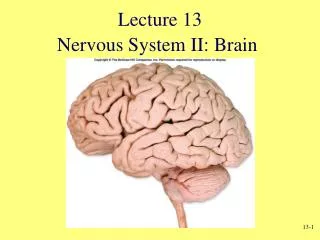
Nervous System II Chapter 12
Major Organs • Brain • Spinal Cord • Cranial Nerves • Spinal Nerves
Brain • May appear somewhat unimpressive at first glance. • Average adult brain- Male 3.5 lbs the Female 3.2 lbs. • Quivering pink tissue with the appearance of cold oatmeal.
Brain • Functions- contains nerve centers associated with sensory functions and is responsible for sensations and perceptions. (motor functions, memory, reasoning, nerve pathways)
Cerebrum • The cerebrum, which develops from the anterior portion of the brain, is the largest part the mature brain. • It consists of two large masses or cerebral hemispheres. • Together they account for about 83% of total brain mass.
A deep bridge of nerve fibers called the corpus callosum connects the cerebral hemispheres. This is the structure that is located beneath the Cerebrum in the pictures. Cerebrum
Many ridges called Gyri or (convolution,) separated by grooves, mark the cerebrum. A shallow to somewhat deep groove is called a sulcus. A very deep grove is called a fissure. The pattern of these elevations and depressions is complex, and it is distinct in all normal brains. Cerebrum
Cerebrum • You need to be able to located the following sulcus and fissures on the brain. • Central Sulcus (Separates the frontal and parietal lobes) • Lateral Sulcus (Separates the temporal lobe from the parietal and frontal lobes) • Transverse Fissure (Is anterior to the cerebellum) • Longitudinal Fissure (Separates the left and right hemispheres)
Cerebrum • Each Cerebral hemisphere has three basic regions • Cortex Matter- A superficial layer that looks gray in fresh brain tissue. • Internal White Matter-internal layer. • Basal Nuclei- islands of gray matter situated deep within the white matter. They relay motor impulses originating in the cerebral cortex and aid in controlling motor activities.
The cerebral cortex is the “executive suite” of the nervous system, where our conscious mind is found. Cerebrum
Cerebrum • A think layer of gray matter called the cerebral cortex, constitutes the outermost portion of the cerebrum. • It covers the gyri or convolutions.
Cerebral Cortex • The cerebral cortex contains nearly 75% of all the neuron cell bodies in the nervous system. • The cerebral cortex has sensory, motor and association areas.
Functions of The Cerebrum • The cerebrum provides higher brain functions: interpreting impulses from sense organs, initiating voluntary muscular movements, storing information as memory, and retrieving this information in reasoning. • The cerebrum is also the seat of intelligence and personality.
Frontal Lobe of the Cerebral • Functions: Motor areas control movements of voluntary skeletal muscles • Association areas carry on higher intellectual processes for concentration, planning complex problem solving, and judging the consequences of behavior.
Parietal Lobe of the Cerebral • Functions: Sensory areas provide sensations of temperature, touch, pressure, and pain involving the skin. • Association areas function in understanding speech and in using words to express thoughts and feelings.
Temporal Lobe of the Cerebral • Functions: Sensory areas are responsible for hearing • Association areas interpret sensory experiences and remember visual scenes, music, and other complex sensory patterns.
Occipital Lobe of the Cerebral • Functions: Sensory areas are responsible for vision. • Association areas combine visual images with other sensory experiences.
Dominant Hemisphere • Both cerebral hemispheres participate in basic functions, such as receiving and analyzing sensory impulses, controlling skeletal muscles on opposite sides of the body. • However, 90% of the population is left hemisphere dominant for the language-related activities of speech, writing, and reading.
Diencephalon • The diencephalon forms the central core of the forebrain and surrounded by the cerebral hemispheres….. contains the thalamus, hypothalamus, and epithalamus.
Thalamus • The thalamus selects incoming sensory impulses and relays them to the cerebral cortex
Hypothalamus • The Hypothalamus is important in maintaining the homeostasis. • Helps maintain the following….Autonomic control center, center for emotional response, body temperature regulation, regulatin of food intake, water intake, and thirst, sleep cycles,etc. • Pituitary Gland- secrets at least 9 hormones. Size and shape of a pea. Located below the hypothalamus. (add to notes)
Epithalamus • The Epithalamus is the most dorsal portion of the diencephalon forms the roof of the third ventricle. • It works in close association with the Hypothalamus and pineal gland. • The Pinal Gland secrets melatonin which plays a role in the sleep/wake cycle. (yes, you have to adjust your notes here)
The Brain Stem • The brain stem extends from the base of the brain to the spinal cord. • The brain stem consists of the midbrain, pons, and medulla oblongata.
The Brain Stem • The midbrain contains reflex centers associated with eye and head movement
The Brain Stem • The pons transmits impulses between the cerebrum and other parts of the nervous system and contains centers that help regulate rate and depth of breathing. • The medulla oblongata transmits all ascending and descending impulses and contains several vital and non-vital reflex centers. (blood pressure, heart rate, and respiration)
Cerebellum • The cerebellum consists of two hemispheres connected by the vermis.
Cerebellum • A thin cortex of gray matter surrounds the white matter of the cerebellum • The arbor vitae (Latin for "Tree of Life") is the cerebellarwhite matter, so called for its branched, tree-like appearance.
Cerebellum • The cerebellum functions primarily as a reflex center, coordinating skeletal muscle movements and maintaining equilibrium.
Optic Chiasma • A point near the thalamus and hypothalamus at which portions of each optic nerve cross over. • We will try to find this in a sheep brain!
Olfactory Bulb • One of two enlargements at the terminus of the olfactory nerve at the base of the brain just above the nasal cavities
Olfactory Bulb • Again this is something that we will really observe in the sheep brain. Larger in sheep than humans!
Cerebrospinal Fluid • The entire surface of central nervous system is bathed by a clear, colorless fluid called cerebrospinal fluid (CSF). • The CSF is contained within a system of fluid-filled cavities called ventricles.
Cerebrospinal Fluid • Found in and around the brain and spinal cord • Reduces brain weight by 97% and prevents the brain from crushing under its own weight. • Protection: the CSF protects the brain from damage by "buffering" the brain. In other words, the CSF acts to cushion a blow to the head and lessen the impact. • Buoyancy: because the brain is immersed in fluid, the net weight of the brain is reduced from about 1,400 gm to about 50 gm. Therefore, pressure at the base of the brain is reduced.
Cerebrospinal Fluid • Excretion of waste products: the one-way flow from the CSF to the blood takes potentially harmful metabolites, drugs and other substances away from the brain. • Endocrine medium for the brain: the CSF serves to transport hormones to other areas of the brain. Hormones released into the CSF can be carried to remote sites of the brain where they may act.
Cerebrospinal Fluid • The CSF is formed by the choroid plexuses that hang from the roof of each ventricle…primarily the first and second ventricles. (lateral) • CSF is replaced in an average human once about every 8 hours.