Download
1 / 2
20 likes | 23 Vues
https://www.creative-biogene.com/blog/index.php/2018/07/29/inhibition-of-the-secretion-of-tumor-exosomes-pd-l1-may-become-an-important-anti-cancer-target-md-anderson-report/

E N D
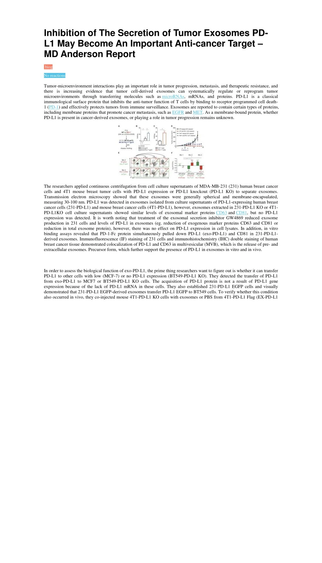
Inhibition of The Secretion of Tumor Exosomes PD- L1 May Become An Important Anti-cancer Target – MD Anderson Report biog No reactions Tumor-microenvironment interactions play an important role in tumor progression, metastasis, and therapeutic resistance, and there is increasing evidence that tumor cell-derived exosomes can systematically regulate or reprogram tumor microenvironments through transferring molecules such as microRNAs, mRNAs, and proteins. PD-L1 is a classical immunological surface protein that inhibits the anti-tumor function of T cells by binding to receptor programmed cell death- 1 (PD-1) and effectively protects tumors from immune surveillance. Exosomes are reported to contain certain types of proteins, including membrane proteins that promote cancer metastasis, such as EGFR and MET. As a membrane-bound protein, whether PD-L1 is present in cancer-derived exosomes, or playing a role in tumor progression remains unknown. The researchers applied continuous centrifugation from cell culture supernatants of MDA-MB-231 (231) human breast cancer cells and 4T1 mouse breast tumor cells with PD-L1 expression or PD-L1 knockout (PD-L1 KO) to separate exosomes. Transmission electron microscopy showed that these exosomes were generally spherical and membrane-encapsulated, measuring 30-100 nm. PD-L1 was detected in exosomes isolated from culture supernatants of PD-L1-expressing human breast cancer cells (231-PD-L1) and mouse breast cancer cells (4T1-PD-L1), however, exosomes extracted in 231-PD-L1 KO or 4T1- PD-L1KO cell culture supernatants showed similar levels of exosomal marker proteins CD63 and CD81, but no PD-L1 expression was detected. It is worth noting that treatment of the exosomal secretion inhibitor GW4869 reduced exosome production in 231 cells and levels of PD-L1 in exosomes (eg. reduction of exogenous marker proteins CD63 and CD81 or reduction in total exosome protein), however, there was no effect on PD-L1 expression in cell lysates. In addition, in vitro binding assays revealed that PD-1-Fc protein simultaneously pulled down PD-L1 (exo-PD-L1) and CD81 in 231-PD-L1- derived exosomes. Immunofluorescence (IF) staining of 231 cells and immunohistochemistry (IHC) double staining of human breast cancer tissue demonstrated colocalization of PD-L1 and CD63 in multivesicular (MVB), which is the release of pre- and extracellular exosomes. Precursor form, which further support the presence of PD-L1 in exosomes in vitro and in vivo. In order to assess the biological function of exo-PD-L1, the prime thing researchers want to figure out is whether it can transfer PD-L1 to other cells with low (MCF-7) or no PD-L1 expression (BT549-PD-L1 KO). They detected the transfer of PD-L1 from exo-PD-L1 to MCF7 or BT549-PD-L1 KO cells. The acquisition of PD-L1 protein is not a result of PD-L1 gene expression because of the lack of PD-L1 mRNA in these cells. They also established 231-PD-L1 EGFP cells and visually demonstrated that 231-PD-L1 EGFP-derived exosomes transfer PD-L1 EGFP to BT549 cells. To verify whether this condition also occurred in vivo, they co-injected mouse 4T1-PD-L1 KO cells with exosomes or PBS from 4T1-PD-L1 Flag (EX-PD-L1
Flag), 4T1-PD-L1KO (EX-PD-L1 KO) into the mammary fat pad of BALB/c mice, then tumors were harvested after 5 days. IF staining of tumor tissue sections showed that EX-PD-L1 Flag instead of EX-PD-L1 KO made 4T1-PD-L1 KO cells positive for PD-L1 Flag. Importantly, the results of flow cytometry analysis further revealed that PD-L1 transported by exosomes is located on the surface of target cells and is capable of binding PD-1. The results indicate that exosomes are capable of transferring functional PD-L1 to other cells. The researchers further examined whether exosomal PD-L1 affects T cell function as exosome PD-L1 binds directly to PD-1. The results showed that exo-PD-L1 significantly inhibited the killing effect of T cells on BT549-PD-L1 KO cells. Furthermore, in order to explore whether exo-PD-L1 inhibits CD3/CD28-triggered T cell activation signaling pathway, they induce PD-1 expression to produce T cell blasts by treating peripheral blood mononuclear cells (PBMC) with phytohemagglutinin (PHA). The results indicate that exo-PD-L1 significantly inhibits ERK phosphorylation and NF-κB activation in CD3/CD28-induced T cells, as well as PHA-induced interleukin-2 (IL-2) secretion, in a dose-dependent manner, all of which are indicators of T cell activation. In addition, exo-PD-L1 from other cancer cell lines, such as colon (RKO) and lung (HCC827) cancer cells, involve similar functions in blocking T cell activation. Collectively, these data support exosomal PD-L1 inhibition of T cell activation. https://www.creative-biogene.com/blog/index.php/2018/07/29/inhibition-of-the-secretion-of-tumor-exosomes-pd-l1-may- become-an-important-anti-cancer-target-md-anderson-report/