Download

1 / 17
190 likes | 774 Vues
Testicular cancer. In a nutshell. It metastasizes early but is highly curable even in metastatic stage Tumor markers important in staging and follow-up. Epidemiology. occurs most often in men between the ages of 15 and 40, where is also the most frequent solid tumor

E N D
In a nutshell • It metastasizes early but is highly curable even in metastatic stage • Tumor markers important in staging and follow-up
Epidemiology • occurs most often in men between the ages of 15 and 40, where is also the most frequent solid tumor • it accounts for only 1% of all cancers in men • incidence highest among men living in the United States and Europe and lowest among men living in Africa or Asia
Risk factors • Undescended testicle=cryptorchidism: -Normally, the testicles descend from inside the abdomen into the scrotum before birth. The risk of testicular cancer is increased in males with a testicle that does not move down into the scrotum -Although most cancers develop in the undescended testicle, about 1 out of 4 cases occur in the normally descended testicle. Based on these observations it is probable that cryptorchidism doesn't actually cause testicular cancer but that there is something else that leads to both testicular cancer and abnormal positioning of one or both testicles. • Family history of testicular cancer: the risk for testicular cancer is greater in men whose brother or father has had the disease.However, only about 3% of testicular cancer cases are actually found to occur in families,most men with testicular cancer do not have a family history of the disease.
Risk factors • Klinefelter's syndrome: Men with Klinefelter's syndrome (a sex chromosome disorder-47XXY-that is characterized by low levels of androgens, sterility, breast enlargement and small testes) are at greater risk of developing testicular cancer. • Race: The risk of testicular cancer among white men is about 5 times that of black men and 3 times that of Asian • Carcinoma in situ of the testicle • A previous testicular cancer
Risk factors • NOT RISK FACTORS: -trauma -repetitive micro-trauma: bicycle or horseback riding
Histology A) From the testicular parenchyma (90%): Germ cell tumors: ~50%/50% seminoma / non-seminomatous germ cell tumor Non-seminomatous germ cell tumors are subdivided in: -Embryonal carcinoma -Yolk sac tumors -Teratoma etc. B) From the testicular stroma (10%): stromal tumors
Routes of dissemination • Local invasion • Lymphatic spread-early -directly to para-aortic lymph nodes (because embryonic origin of testes is intra-abdominal) • Hematogenous metastases-early -lung, liver, brain, bone
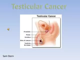
Clinical picture • Most patients present with symptoms consistent with infectious epididymitis and/or orchitis • only a minority of patients present with the pathognomonic painless testicular mass • Lumbar pain-in case of para-aortic lymph node involvement • Gynecomastia • Stromal tumors may secrete estradiol directly • βHCG secreted by the tumor acts like LH and increases testosterone, which transforms in adipose tissue into estradiol • Symptoms caused by metastases
Diagnosis • History and physical • Inspection and palpation of the testicle • Testicular ultrasound • Palpation of inguinal lymph nodes • Abdominal CT for para-aortic and hepatic metastases • Tumor markers (βHCG, AFP, LDH), VSH • Chest radiography for lung metastases • Chest CT for lung metastases IF para-aortic lymph nodes positive or chest radiograph abnormal • Brain MRI for brain metastases IF clinical signs of brain metastases • Bone scintigraphy IF bone pain βHCG=human chorionic gonadotropin, beta subunit AFP=alpha-fetoprotein LDH=lactate dehydrogenase
Diagnosis-tumor markers • Seminoma: AFP negative!!!,βHCG may be positive, LDH may be increased • Non-seminomatous germ cell tumor: AFP, βHCG may be positive; LDH may be increased Mnemonic: AFP-”fetal tissue growing in the testis” in case of non-seminoma
Treatment • If suspect for testicular cancer=>UNILATERAL RADICAL INGUINAL ORCHIECTOMY • NO BIOPSY BECAUSE THE RISK OF DISSEMINATION !!! Radical orchiectomy. Note the inguinal approach and the use of the tourniquet.
Adjuvant treatment for seminoma Stage I (=no lymphadenopathy on CT and no distant metastases): Choice between: -surveillance -one cycle of chemotherapy -para-aortic (“prophylactic”) radiotherapy (low dose: 25-30 Gy) Stage II (=positive lymphadenopathy) Choice between: • para-aortic plus ipsilateral inguinal radiotherapy • 4 cycles of chemotherapy Stage III (=distant metastases or highly elevated tumor markers) -4-6 cycles of chemotherapy
Adjuvant treatment for non-seminomatous germ cell tumor -para-aortic lymph node dissection or chemotherapy (more complicated guidelines, too much for student level)
Follow-up • History and physical + chest X-ray + βHCG, AFP, LDH -every 2 months for year 1 • 3 mo/y 2 • 4 mo/y 3 • 6 mo/y 4 • Abdominal CT after 4 months of treatment • PET-CT if suspicion of relapse
Follow-up for side effects of treatment • RT favorizes gastric cancer • RT may lead to retroperitoneal fibrosis with ureteral obstruction • Chemotherapy may lead to sterility (although in general men with testicular cancer have low fertility from the beginning due to degenerative changes in the contralateral testicle) Etc.