Testicular Cancer
140 likes | 1.38k Vues
Testicular Cancer. Dr. Belal M. Hijji, RN. PhD May 30, 2011. Learning Outcomes. By the end of this lecture, students will be able to: Recognise the prevalence and classification of testicular cancer, as well as risk factors and clinical manifestations

Testicular Cancer
E N D
Presentation Transcript
Testicular Cancer Dr. Belal M. Hijji, RN. PhD May 30, 2011
Learning Outcomes By the end of this lecture, students will be able to: • Recognise the prevalence and classification of testicular cancer, as well as risk factors and clinical manifestations • Gain insight into assessment, diagnostic findings, and medical management of Ca testis • Discuss nursing management of pt with Ca testis
Testicular Cancer • Is the most common cancer in men 15 to 35 years although it is rare (up to 10.3 cases per 100, 000) • Can occur in males of any age. • Is a highly treatable and usually curable form of cancer. • The testicles contain several types of cells, each of which may develop into one or more types of cancer. • The type of cancer determines the appropriate treatment. • Testicular cancers are classified as secondary, germinal or nongerminal.
Types of Testicular Cancer • Germinal Tumors • Over 90% of all cancers of the testicle are germinal and these may be further classified as seminomas or nonseminomas based on histology. • Seminomas develop from the sperm-producing cells of the testes and tend to be localised, whereas nonseminomatous tumors grow quickly.
Nongerminal Tumors • Testicular cancer may also develop in the supportive and hormone producing tissues, or stroma, of the testicles. • The two main types of stromal tumors are Leydig cell tumors and Sertoli cell tumors. • These tumors infrequently spread beyond the testicle. However, a small number of them metastasize and tend to be resistant to chemotherapy and radiation therapy.
Secondary Testicular Tumors • These tumors have metastasized to the testicle from other organs. Lymphoma is the most common cause of secondary testicular cancer. • The prognosis for these cancers is usually poor. • Treatment depends on the specific type of cancer
Risk Factors • Men with any type of undescended testis are at greater risk than in the general population. • Family history of testicular cancer and cancer of one testicle. • Race and ethnicity: Caucasian American men have a five times greater risk than that of African American. • Occupational hazards, including exposure to chemicals encountered in mining, are potential risk factors. • Prenatal exposure to DES (Diethylstilboestrol). This synthetic nonsteroidal estrogen was given from about 1940 to 1970 to pregnant women under the mistaken belief it would reduce the risk of pregnancy complications and losses.
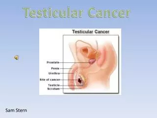
Clinical Manifestations • A mass or lump on the testicle and generally painless enlargement of the testis. • Possible heaviness in the scrotum, inguinal area, or lower abdomen. Backache (from retroperitoneal node extension), abdominal pain, weight loss, and general weakness due to metastasis. • Enlargement of the testis without pain is a significant diagnostic finding. Testicular tumors tend to metastasize early, spreading to the lymph nodes in the retroperitoneum and to the lungs.
Assessment and Diagnostic Findings • Monthly testicular self-examinations (TSEs). • Possible elevation of human chorionic gonadotropin and alpha-fetoprotein in blood. • Intravenous urography to detect any ureteral deviation caused by a tumor mass. • Lymphangiography to assess the extent of tumor spread to the lymphatic system. • Ultrasound to determine the presence and size of the testicular mass. • CT scan of the chest, abdomen, and pelvis to determine the extent of metastasis • Biopsy is the only definitive diagnostic modality.
Medical Management • The goals of management are to eradicate the disease and achieve a cure. • The testis is removed through an inguinal incision with a high ligation of the spermatic cord. • The patient may develop ejaculatory dysfunction with resultant infertility. Thus, sperm banking before surgery may be considered. • Postoperative irradiation of the lymph nodes is used in treating seminomas. • Radiation is delivered only to the affected side and is indicated in situations where the disease does not respond to chemotherapy or for whom lymph node surgery is not recommended.
Testicular carcinomas are highly responsive to chemotherapy. • Combining different types of treatment, including surgery, radiation therapy, and chemotherapy.
Nursing Management • Assessment of the patient’s physical and psychological status • Monitoring for response to and possible effects of surgery, chemotherapy, and radiation therapy. • Issues related to body image and sexuality are addressed. • Teaching the patient that radiation therapy will not necessarily prevent him from fathering children, nor does unilateral excision of a testis necessarily decrease virility. • Reminding the patient about the importance of performing TSE and keeping follow-up appointments with the physician.