Testicular Pathology
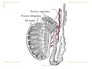
Testicular Pathology. Emad Raddaoui , MD, FCAP, FASC Associate Professor & Consultant. Normal Anatomy. Testicular diseases. Epididymitis And ORCHITIS : Inflammatory conditions are generally more common in the epididymis than in the testis.


Testicular Pathology
E N D
Presentation Transcript
Testicular Pathology EmadRaddaoui, MD, FCAP, FASC Associate Professor & Consultant
Testicular diseases Epididymitis And ORCHITIS: • Inflammatory conditions are generally more common in the epididymis than in the testis. • However, some infections, notably syphilis, may begin in the testis with secondary involvement of the epididymis • Epididymitis and possible subsequent orchitis are commonly related to infections in the urinary tract (cystitis, urethritis, genitoprostatitis) • These infections reach the epididymis/testis through either the vas deference or the lymphatics of the spermatic cord
Epididymitis CAUSES: Varies with age • Children: uncommon, usually associated with a congenital genitourinary abnormality and infection with Gram –ve rods. • In sexually active men < 35 years Chlamydia trachomatis and Neisseria • Older than 35 Years E.Coli and Pseudomonas.
Epididymitis And Orchitis Microscopic findings: • Non specific acute inflammation characterized by congestion, edema and infiltration by lymphocytes, neutrophilsand macrophages. • Initially involves the interstitial connective tissue later involves tubules may progress to frank abscess. • Often followed by fibrous scarring. • Leydig cells are not usually destroyed
Granulomatous (Autoimmune) Orchitis • Usually middle–aged men, unilateral testicular mass. • Moderately tender but sometimes may present as painless testicular mass; mimicking a testicular tumor. • Although an autoimmune basis is suspected, the cause of these lesions remain unknown. • May be a response to acid-fast products of disintegrated sperm, post-infectious, or due to trauma or sarcoidosis • Microscopically: granulomas, restricted within the spermatic tubules.
Specific Inflammations: Gonorrhea: Extension of infection from the posterior urethra prostate seminal vesicles epididymis is the usual course of a neglected gonococcal infection. • Can lead to frank abscess may spread to testis and can produce a supurativeorchitis. Tuberculosis: • Almost invariably begins in the epididymis and may spread to the testis. • In many of these cases ,there is associated tuberculousprostatitis and seminal vesiculitis • Microscopy: CaseatingGranulomatous inflammation.
* Pink Leyding cells are seen here in the interstitium( arrow). Note the pale golden brown pigment as well( bold arrow). There is active spermatogenesis (*)
Testicular Tumors • Complex mixture of anatomic types • 95% of them originate from germ cells, • Age group15-30 years, whites> blacks • Most of gem cell tumors are highly aggressive cancers • Capable of wide ,extensive dissemination • Current therapy ,most of them can be cured • Non germinal tumors are generally benign
Testicular TumorsClassification • Germ cell tumors : Seminomatous: • Seminoma • Spermatocyticseminoma Non Seminomatous • Embryonal carcinoma • Yolk sac (endodermal Sinus) tumor • Choriocarcinoma • Teratoma • Sex Cord Tumors • Leydig cell tumor • Sertoli cell tumor
Testicular Tumors Pathogenesis • Predisposing factors: -Cryptorchidism :10% of testicular tumors -Testicular dysgenesis -Genetic factors
Seminoma • The most common type of germ cell tumors (50%) • Peak incidence in thirties (Almost never occur in infants) • Identical one occurs in the ovary (Dysgerminoma)
Bulky masses • Homogenous • Gray-white • Lobulated cut surface • Usually no necrosis or hemorrhage • In 50% ,the entire testis is involved • Occasionally extends to the epididymis, spermatic cord, or scrotal sac Seminoma
Seminoma of the testis appears as a fairly well-circumscribed, pale, fleshy, homogeneous mass
Seminoma , Morphology • Microscopically ,sheets of uniform cells • Lobules separated by delicate fibrous septa with many lymphocytes • Cells are large ,round ,has distinct cell membrane • Large nucleus with prominent nucleolei • Positive for PLAP
Sheets of uniform cells • Lobules separated by delicate fibrous septa with many lymphocytes • Cells are large ,round ,has distinct cell membrane • Large nucleus with prominent nucleolei • Positive for PLAP
Large cells with distinct cell borders, pale nuclei, prominent nucleoli, and a sparse lymphocytic infiltrate
SpermatocyticSeminoma • Distinctive tumor ,clinically and histologically • 1-2 % of testicular tumors • Over age 65 • Slow growing tumor ,rarely metastasise • Prognosis is excellent
Embryonal Carcinoma • 20 to 30 year age group • More aggressive than seminomas • Smaller than seminoma • Grossly, shows foci of necrosis and hemorrhage • Microscopically,showssheets of undifferentiated cells as well as primitive glandular differentiation. Cells grow in alveolar or tubular pattern ,sometimes with papillary convolutions. • Could be present with other neoplasm in 45%
Embryonalcarcinoma hemorrhagic mass.
Embryonal carcinoma shows sheets of undifferentiated cells as well as primitive glandular differentiation. The nuclei are large and hyperchromatic
Yolk Sac Tumor • Also known as Endodermal sinus tumor • The most common tumor in infant and children up to 3 years of age • Has a very good prognosis • Non encapsulated ,homogenous ,mucinousappearance
Mixed germ cell tumor of testes, with embryonal carcinoma, yolk sac tumor
Yolk Sac Tumor • Microscopically ,structures resemble endodermal sinuses • Schiller-Duval bodies • Hyaline –pink globules • AFP positive
Schiller-Duval body is a structure seen in yolk sac tumor. It consists of a central vessel surrounded by tumor cells – the whole structure being contained in a cystic space often lined by flattened tumor cells
An endodermal sinus tumor (yolk sac tumor) of the testis is shown composed of primitive germ cells that form glomeruloid or embryonal-like structures. These tumors are most frequent in children, but overall they are rare.
Choriocarcinoma • Highly malignant tumor • Cytotrophoblastic and syncytiotroblasticcells • Small lesions • HCG positive
Teratoma • Various cellular or organoid components • Any age ,infancy to adult life • Mature forms are common in infants and children • Adult forms are rare • As a component with other type in 45%
Teratoma • Usually large 5 -10 cm • Heterogeneous appearance • Hemorrhage and necrosis indicate embryonal component • Composed of heterogenous collection of cells or organoid structures • Neural tissue ,cartilage ,squamous epithelium ,glandular components….
Teratoma • Germ cell tumors could arise from teratoma • In children ,mature teratomas behave benign • In post pubertal male ,all teratomas regarded malignant ,and capable of metastasis ,regardless of whether the elements are mature or not.
A small testicular carcinoma is shown here. There is a mixture of bluish cartilage with red and white tumor tissue. This neoplasm microscopically contained mainly teratoma, but areas of embryonal carcinoma were also present
At the bottom is a focus of cartilage. Above this is a primitive mesenchymalstroma and to the left a focus of primitive cells most characteristic for embryonal carcinoma. This is embryonal carcinoma mixed with teratoma.
Testicular tumorsClinical Features • Biopsy of a testicular tumor is associated with a risk of tumor spillage • The standard management of solid tumors is radical orchiectomy • Lymphatic spread is common • Retroperitoneal and para-aortic nodes are first to be involved • Hematogenous spread to Lung, liver, Brain, and bones.