Download

1 / 26
320 likes | 737 Vues
Testicular Neoplasms. - In the 15- to 34-year-old age group, they are the most common tumors of men. - Tumors of the testis are a heterogeneous group of neoplasms that include: I. Germ cell tumors : 95%; all are malignant.

E N D
Testicular Neoplasms - In the 15- to 34-year-old age group, they are the most common tumors of men. - Tumors of the testis are a heterogeneous group of neoplasms that include: I. Germ cell tumors : 95%; all are malignant. II. Sex cord-stromal tumors: from Sertoli or Leydig cells; usually benign. Note: The cause of testicular neoplasms remains unknown
Risk factors: 1. more common in whites than in blacks. 2. Cryptorchidism 3- to-5 folds increase in the risk of cancer in the undescended testis, and an increased risk of cancer in the contralateral descended testis. 3. Intersex syndromes a. Androgen insensitivity syndrome b. Gonadaldysgenesis 4. Family history Brothers of males with germ cell tumors have an 8-10-fold increased risk over that of the population
5. The development of cancer in one testis markedly increased risk of neoplasia in the contralateral testis. 6. An isochromosome of the short arm of chromosome 12, i(12p), is found in virtually all germ cell tumors, regardless of their histologic type. Note: - Most testicular tumors in postpubertal males arise from the in situ lesion intratubular germ cell neoplasia; - This lesion is present in conditions associated with a high risk of developing germ cell tumors (e.g., cryptorchidism, dysgenetic gonads
Testicular germ cell tumors are subclassified into: I. Seminomas II. Non-seminomatous germ cell tumors(NSGCT) - The histologic appearances may be: 1. Pure (i.e., composed of a single histologic type 60% of cases) 2. Mixed (seen in 40% of cases).
I. Seminomas, - Make up 30-40% of all testicular tumors - They are divided into two major categories: a. Classic seminoma • Spermatocyticseminoma Classic seminoma - 90% of the cases - fourth decade of life - rare in prepubertal children - progressive painless enlargement of the testis - histologically identical to ovarian dysgerminomas and to germinomas occurring in the CNS and other extragonadal sites.
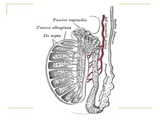
MORPHOLOGY Gross - soft, well-demarcated tumors - usually without hemorrhage. Microscopically • The cells are large and uniform with distinct cell borders; Have clear, glycogen-rich cytoplasm; round nuclei with nucleoli. They are arranged in small lobules with intervening fibrous septa. A lymphocytic infiltrate is usually present along with an ill-defined granulomatous reaction
Spermatocyticseminoma - Should be separated from classic seminoma - comprises 10% of all cases of seminoma - Is a distinct clinical and histologic entity. - Is an uncommon tumor • It occurs in much older individuals (older than 65 years of age.) Microscopically - Composed of cells with round nuclei and prominent variation in size - The cytoplasm is dense and devoid of glycogen
- In contrast with classic seminomas, spermatocyticseminomas: a. Lack lymphocytic infiltrates, granulomas, and syncytiotrophoblasts • Are not admixed with other germ cell tumor histologies c. Are not associated with intratubular germ cell neoplasia; d. More commonly bilateral e. Excellent prognosis with no metastasis f. Do not occur in extratesticular sites
2. Embryonal carcinomas : • Are ill-defined, invasive masses containing foci of hemorrhage and necrosis Microscopically The tumor cells are large and primitive-looking. The cytoplasm is basophilic. The neoplastic cells may be arranged in solid sheets or as primitive glandular structures and papillae Note • In most cases, other germ cell tumors (yolk sac tumor, teratoma, choriocarcinoma) are admixed with the embryonalca. - Pure embryonal carcinomas account for only 2% to 3% of all testicular germ cell tumors
3. Yolk sac tumors - Are the most common primary testicular neoplasm in children younger than 3 years of age and in this age group it has a very good prognosis. • In adults, yolk sac tumors most often are seen admixed with embryonal carcinoma, with a worse prognosis • In nonseminomatous germ cell tumors, the incidence of yolk sac elements is 80%
Histologic examination - The tumor is composed of low cuboidal to columnar epithelial cells forming Microcysts, Lacelike (reticular) patterns and Sheets, Glands, and papillae . A distinctive feature is the presence of structures resembling primitive glomeruli, called Schiller-Duvall bodies. - have eosinophilic hyaline globules in which α1-antitrypsin and alpha fetoprotein (AFP) can be demonstrated by immunohistochemical techniques. - AFP can also be detected in the serum.
4. Choriocarcinomas - the germ cells differentiate along trophoblastic lines. - about 5% of all testicular tumors Grossly - primary tumors may be small lesions, even those with extensive systemic metastases • May show total necrosis and tumor regression and is the most common tumor that undergoes regression Microscopic examination • composed of sheets of small cuboidal cells (cytotrophoblasts) irregularly intermingled with large, eosinophilicsyncytial cells containing multiple dark, pleomorphic nuclei(syncytiotrophoblasts). • HCG within syncytiotrophoblasts can be identified by immunohistochemical staining and is elevated in the serum
5. Teratomas - the neoplastic germ cells differentiate along somatic cell lines. -Gross: - These tumors form firm masses that on cut surface often contain cysts and areas of cartilage. - They may occur at any age from infancy to adult life. • Pure forms of teratoma are common in infants and children , being second in frequency only to yolk sac tumors - In adults, pure teratomas are rare, constituting 2% to 3% of germ cell tumors, most are seen in combination with other histologic types
Microscopically 1. Mature teratomas a heterogeneous, collection of differentiated cells or organoid structures, such as neural tissue, musclebundles, islands of cartilage, clusters of squamous epithelium, thyroid tissue, bronchial epithelium, and of intestinal wall or brain substance, all embedded in a fibrous or myxoidstroma 2. Immature teratoma • Sharehistologic features with fetal or embryonal tissues 3. Teratoma with malignant transformation
Clinical Features of testicular germ cell neoplasms - present most frequently with a painless testicular mass (enlargement is non-translucent). - Biopsy of a testicular neoplasm is associated with a risk of tumor spillage, which would necessitate excision of the scrotal skin in addition to orchiectomy. - standard management of a solid testicular mass is radical orchiectomy, based on the presumption of malignancy
- Seminomas and nonseminomatous tumors differ in their behavior and clinical course: I. Seminomas a. Often remain confined to the testis for long intervals and may reach considerable size before diagnosis. b. Metastases most commonly are encountered in the iliac and paraaortic lymph nodes c. Hematogenous metastases occur late in the course of the disease
II. Nonseminomatous germ cellneoplasms a. Tend to metastasize earlier, by lymphatic as well as hematogenous routes. b. Hematogenous metastases are most common in the liver and lungs. c. Metastatic lesions may be identical to the primary testicular tumor or may contain elements of other germ cell tumors.
Assay of tumor markers secreted by germ cell a. These markers along with some clinical and morphologic features are helpful diagnostically b. have an even more valuable role in following the response of tumors to therapy after the diagnosis is established. I. Human chorionic gonadotropin (hCG) - Is always elevated inchoriocarcinoma. 2. Increased alpha fetoprotein (AFP) indicates a yolk sac tumor component. 3. The levels of lactate dehydrogenase (LDH) correlate with the tumor burden.
The treatment of testicular germ cell neoplasms I. Seminoma: radiosensitive and tends to remain localized for long periods, has the best prognosis (95% of patients with early-stage disease can be cured) II. nonseminomatous germ cell tumors, the histologic subtype does not influence the prognosis significantly, and hence these are treated as a group. • 90% of the patients achieve complete remission with aggressive chemotherapy, and most are cured. - Pure choriocarcinoma carries a dismal prognosis. - With all testicular tumors, recurrences, typically in the form of distant metastases, usually occur within the first 2 years after treatment