Testicular Pathology
270 likes | 593 Vues
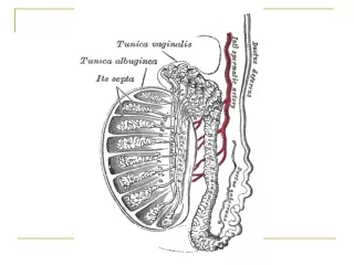
Testicular Pathology. Pathology Department KSU, Riyadh March 2018 Reference: Robbins & Cotran Pathology and Rubin’s Pathology. Lecture outline. At the end of this lecture, the student should be able to: Have a working knowledge of the normal histology of the testis and epididymis.

Testicular Pathology
E N D
Presentation Transcript
Testicular Pathology Pathology Department KSU, Riyadh March 2018 Reference: Robbins & Cotran Pathology and Rubin’s Pathology
Lecture outline At the end of this lecture, the student should be able to: • Have a working knowledge of the normal histology of the testis and epididymis. • Know the predisposing factors and pathology of epididymitis. Epididymitis and orchitis • Non specific Epididymitis and orchitis • Granulomatous/Autoimmune Orchitis • Gonorrhea • Tuberculosis • Be familiar with the basic classification and pathology of testicular tumors. Testicular tumors • seminoma • yolk sac tumor • embryonal carcinoma • Teratoma • choriocarcinoma.
Taken from Robin and Cotran pathological basis of disease 2010 by Saunders,
Testicular diseases Epididymitis and orchitis: • Epididymitis: inflammation of epididymis • Orchitis: inflammation of testis • Inflammatory conditions are generally more common in the epididymis than in the testis. • However, some infections, notably syphilis, may begin in the testis with secondary involvement of the epididymis
Inflammation: epididymitis and orchitis 1.Non specific epididymitis and orchitis: • are commonly related to infections in the urinary tract (cystitis, urethritis and genitoprostatitis). • infections reach the epididymis/testis through the vas deference or the lymphatics of the spermatic cord. • Causative organisms vary with age; • Children: it is uncommon. Usually associated with a congenital genitourinary abnormality and infection with Gram –ve rods. • In men younger than age 35 years: Chlamydia trachomatis and Neisseria are common causative organisms. • In men older than 35 Y: E.Coliand Pseudomonas. • Microscopic findings: • congestion, edema and infiltration by neutrophils, macrophages and lymphocytes. • initially involves the interstitium but later involves seminiferous tubules • may progress to frank abscess. • Heals by fibrous scarring. • Leydig cells are not usually destroyed.
Inflammation: epididymitis and orchitis 2.Granulomatous (autoimmune) epididymitis & orchitis: • middle–aged men present with unilateral testicular mass. • mimic testicular tumor. • microscopy: granulomatous inflammation with plasma cells and lymphocytes. • autoimmune basis is suspected. • May be in response to disintegrated sperm, post-infectious, due to trauma or sarcoidosis. 3.Gonorrhea: • Gonoccocal infection can spread from urethra to prostate, seminal vesicles and then to epididymis and testis leading to suppurativeorchitis and even abscess. 4.Tuberculosis: • Begins in the epididymis and spreads to the testis. • There is associated tuberculous prostatitis and seminal vesiculitis • Microscopy: CaseatingGranulomas
Testicular Tumors • Testicular tumors are the most important cause of firm, painless enlargement of testis. • Peak incidence between the ages of 20 and 34 years.
Classification of testicular tumors Testicular tumors are a heterogeneous group of tumors divided into germ cell tumors and sex cord stromal tumors: 1) GERM CELL TUMORS (95% of testicular tumors) • Tumors with One Histologic Pattern (pure form) • Seminomatous germ cell tumors: • Seminoma • Spermatocytic seminoma • Non-Seminomatous germ cell tumors (NSGCT): • Embryonal carcinoma • Yolk sac (endodermal Sinus) tumor • Choriocarcinoma • Teratoma: they can be mature, immature or with malignant transformation • Tumors with more than one Histologic Pattern: mixed germ cell tumor (mixed form) 2) SEX CORD STROMAL TUMORS. • Leydigcell tumor • Sertolicell tumor • In adults, 95% of testicular tumors are germ cell tumors, and all are malignant. • Sertolior Leydig cells (sex cord/stromal tumors) are uncommon and are usually benign.
GERM CELL TUMORS (GCT) • Between 15-30 years of age, these are the most common tumor in men. • Most of gem cell tumors are highly aggressive cancers, capable of extensive dissemination • Good news is that with current therapy most of them can be cured. • Germ cell tumors may have • a single tumor type component • or as is 60% of cases a mixture of tumor types e.g. mixtures of seminomatous and non-seminomatous components. • Most GCTs originate from precursor lesions called intratubular germ cell neoplasia (it is like carcinoma-in-situ)
GERM CELL TUMORS (GCT) Predisposing factors: • Cryptorchidism is associated with a 3 to 5 fold increase in the risk of cancer in the undescended testis and in the contralateral descended testis. About 10% cases of testicular cancer have cryptorchidism. • Testicular dysgenesis • Genetic factors • Strong family predisposition. Brothers, fathers and sons of testicular cancer patients are at risk. • There is a high risk of developing cancer in one testis if the contralateral testis has cancer. • Testicular tumors are more common in whites than in blacks.
Seminoma • Is the most common type of testicular tumors. • It is also the most common type of testicular GCT (50%) • Almost never occur in infants • Peak incidence in the 30ies • An identical tumor occurs in the ovary (called dysgerminoma). • Classic seminoma is highly sensitive to radiation therapy, and the overall 5-year survival is 90 to 95%. • Gross morphology • Bulky masses, sometimes very large • Homogenous ,gray-white, lobulated cut surface • No necrosis or hemorrhage Taken from Robin and Cotran pathological basis of disease, 2010 Saunders
Seminoma Microscopic morphology • sheets of uniform cells divided into lobules by delicate fibrous septa containing lymphocytes. • Cells are largeand round with large nucleus and prominent nucleoli • Cytoplasm of tumor cell has glycogen • Positive for PLAP, OCT4 stain and c-kit (CD117). Taken from Robin and Cotran pathological basis of disease, 2010 Saunders
SpermatocyticSeminoma • Uncommon: 1-2 % of testicular GCTs • Over age 65 • Slow growing tumor, does not metastasize • Prognosis is excellent
Embryonal Carcinoma • It accounts for about 15 to 35% of testicular GCTs • 20 to 30 year age group • More aggressive than seminomas • metastasizes early via both lymphatic and hematogenousroutes. • Radiation is not as effective as with seminoma, but newer chemotherapeutic agents are very effective with greatly improved prognosis. • Grossly: smaller than seminoma, poorly demarcated • Variegated with foci of necrosis and hemorrhage • Can be seen combined with other GCTs (in mixed GCTs) • Tumor cells are positive for cytokeratin (CK) and CD30 stain http://www.humpath.com/spip.php?article4540
Yolk Sac Tumor • Also called Endodermal sinus tumor • Testicular yolk sac tumors occur in two forms: • as a pure form in young children (pure YST of the adult testis is rare) • as in combination with other NSGCTs, mainly embryonal carcinoma, in adults. • It is the most common tumor in infant and children up to 3 years of age and it has a very good prognosis in infants and children. • In adults it occurs as a part or component of mixed GCT (commonly mixed with embryonal carcinoma) • Patients have elevated serum alpha fetoprotein (AFP). AFP may be used as a marker of disease progression in the patient's serum and also aid in diagnosis. • The biologic behavior of YST is similar to that of embryonal carcinoma • Gross morphology: • Non encapsulated, homogenous, yellow white, mucinous • Microscopic morphology • Tumor shows structure resembling endodermal sinuses called as Schiller-Duval bodies are characteristic. • Hyaline–pink globules • Tumor cell are positive for alphafetoprotein (AFP) and alpha-1-antitrypsin stain.
Choriocarcinoma • Highly malignant tumor • Patients have elevated serum human chorionic gonadotropin (HCG) • Small sized lesions • Prominent hemorrhage and necrosis • Made up of malignant trophoblastic (placental) tissue (cyto-trophoblastic and syncytio-troblastic cells) • Tumor cells positive for human chorionic gonadotropin (HCG) stain • Pure choriocarcinoma of the testis is extremely rare, and the tumor is much more common as a component of mixed GCT.
Teratoma • It is a tumor composed of various different types of cells or organ components • Any age, infancy to adult life • In its pure form it is common in infants and children second to yolk sac tumor (in this age group) • In adult the pure form is rare. It occurs as part of mixed GTC webpath.med.yale.edu www.humpath.com
Teratoma • Usually large 5 -10 cm • Heterogenous appearance with solid and cystic areas. Can show bone, cartilage and teeth grossly. • Composed of bizzarely distributed collection of different type of cells or organ structures (heterogenous) • Any of the following cell types of various organs can be present: neural/brain, cartilage, bone, squamous epithelium, hair, glandular cells, smooth muscle, thyroid tissue, bronchial epithelium of lung, pancreatic tissue etc. • If the cells/tissue is mature looking it is called as mature teratoma. • If some of the cells/tissue component is immature it is called as immature teratoma. • If any of the cells/tissue undergoes non germ cell type of malignant tranformation it is called as teratoma with malignant transformation (rare) e.g squamous cells develop squamous cell carcinoma or the glandular cells develop adenocarcinoma. • Behavior of teratomas: • In infants and children, mature teratomas are benignand immature teratoma is considered malignant. • In post pubertal male, all teratomas are regarded as malignant, and capable of metastasis, regardless of whether the elements are mature or not.
www.auanet.org/education/modules/pathology/testis-germ/teratoma.cfmwww.auanet.org/education/modules/pathology/testis-germ/teratoma.cfm
Mixed GCTs • Mixed Germ Cell Tumors are quite common. • About half of testicular tumors are composed of a mixture of GCTs. • The common combinations/mixtures are: • Teratoma + embryonal carcinoma +/- yolk sac tumor • Seminoma + embyronal carcinoma
Clinical features of GCTs • Present as a painless enlarging mass in the testis. Generally any solid testicular mass should be considered neoplastic. • Germ cell tumors secrete hormones and enzymes that can be detected in blood (HCG, AFP, and lactate dehydrogenase) • Biopsy of a testicular tumor is associated with a risk of tumor spillage therefore it is not recommended. • The standard management of solid testicular tumors is radical orchiectomy • GCTs can spread by direct extension to the epididymis, spermatic cord, or scrotal sac • Lymphatic spread is common. Retroperitoneal and para-aortic nodes are first to be involved • Hematogenous spread to Lung, liver, Brain, and bones. • Seminomatous tumors are radiosensitive. • Non-seminomatous tumors are chemosensitive and respond very well to chemotherapy.
Prognosis • More than 95% of patients with seminoma can be cured • 90% of patients with non-seminomatoustumorscan achieve complete remission with aggressive chemotherapy, and most can be cured • The rare pure choriocarcinoma is the most aggressive non-seminomatous tumor. Pure choriocarcinoma has a poor prognosis
Difference between seminoma and non-seminomatous germ cell tumors