Download
1 / 71
760 likes | 1.38k Vues
Testicular Cancer. Adrian Clubb Greenslopes Hospital. References. Smith’s General Urology Campbells Urology European Urology (March 08):

E N D
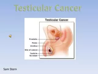
Testicular Cancer Adrian Clubb Greenslopes Hospital
References • Smith’s General Urology • Campbells Urology • European Urology (March 08): • European Consensus Conference on Diagnosis and Treatment of Germ Cell Cancer: A Report of the Second Meeting of the European Germ Cell Cancer Consensus group (EGCCCG): Part I
Overview • Most common solid tumour in men 15 – 30 years • Incidence ~1/1,600 (exact figure varies on source) • Right > left (cryptochidism more common on right) • Bilateral in 1-2%
Aetiology • Cryptochidism represents ~10% of cases • Risk 1/20 intra-abdominal, 1/80 inguinal • Exogenous estrogens to mother • Atrophy (nonspecific or Mumps related) • Trauma/infection – not proven
Staging - TNM • T • 1 – limited to testis and epididymis, no vascular invasion • 2 – invades beyond tunica albuginea or has vascular invasion • 3 – invades spermatic cord • 4 – invades scrotum
N • 1 - lymph node metastasis <2cm and <5 nodes • 2 – metastasis in >5 nodes, nodal mass 2-5cm • 3 – nodal mass >5cm • M • 1 – distant metastasis present
Staging – AJCC (American Joint Comittee on Cancer) • Stage 0 – CIS • Stage I – T1-4/N0/M0 • IA – T1 • IB – T2-4 • IS – ANY T, S1-3 • Stage II – T1-4/N1-3/M0 • IIA – N1 • IIB – N2 • IIC – N3 • Stage III – T1-4/N1-3/M1
Presentation • Painless enlargement/mass of testis • Acute pain ~ 10% (intratesticular haemorrhage or infarction) • Metastatic disease ~10% • Following trauma (incidentaloma)
Firm, nontender mass • Hydrocoele • Abdominal mass with advanced retroperitoneal disease • Gynaecomastia ~5% GCT (but 30-50% Sertoli/Leydig tumours)
Initial Investigations • Tumour Markers (ensure prior to surgery) • CT Abdomen/Pelvis • CXR • Testicular US (often done but should never delay surgery)
Tumour Markers • Alpha-fetoprotein • Trophoblasts • Major serum binding protein produced by foetal yolk sac, liver, GIT • Negligible amounts after 1 year of age • Half – life 4-6 days • Beta-human chorionic gonadotrophin • Syncytiotrophoblasts • Secreted by placenta for maintanence of corpus luteum • Half – life 24 hours • LDH • Correlates with tumour burden
Surgery • Inguinal orchidectomy • High vascular ligation (internal ring) • Frozen section only if diagnosis in doubt or for organ sparing • Prolene stitch tie to cord with long tail (in order to find later if RPLND necessary)
Surgical Pitfalls • Dont forget fertility issues – sperm banking an option • Haemorrhage most common complication • Acute painful scrotal swelling • Retroperitoneal bleed • Bleeding: • Testicular artery • Cremesteric branch • Scrotal (gubernaculum)
Scrotal approach • Cannot ligate the cord high enough • Higher local recurrence rate • Never biopsy
Fertility Issues • ~25% have fertility issues at time of presentation • High concentrations of anti-sperm antibodies than general population • ~50% subfertile post-orchidectomy • Further attacks on fertility common (RPLND, chemo, radio) • 35% pregnancy rate in one US study • Offer sperm banking prior to chemotherapy (Europeans recommend offering prior to orchidectomy)
Pathology • GCT • Seminoma • Classical • Anaplastic • Spermatocytic • Non-Seminoma • Embryonal • Yolk sac • Teratoma • Choriocarcinoma • Mixed
CIS • Non GCT • Leydig • Sertoli • Gonadoblastomas • Secondary • Lymphoma (most common testicular malignancy in >50 year olds) • Leukemic infiltration • Metastatic
Seminoma • Three histologic subtypes • Classical – 85% (3rd decade) • Anaplastic – 5-10%, metastasis early • Spermatocytic – 5-10% (>50 year olds), v. Good prognosis • Tumour markers often normal, B-HCG can be raised (but <500) • Overall survival >99%
Prognostic Indicators • Size of tumour (>4cm) • Infiltration of the rete testis • Any positive tumour markers
Treatment • Stage I (T1-4/N0/M0) • 32% will relapse if 2 or more risk factors • 12% relapse with no risk factors • 97% of relapses in retroperitoneal and iliac lymph nodes • Relapse can occur as late as 10 years
Surveillance • Radiotherapy • Retroperitoneum • Retroperitoneum and iliac nodes • Chemotherapy (not favoured in Australia)
Surveillance • Up to 88% “cured” by orchidectomy alone • Patient needs to be reliable • Psychological stress potential negative (difficult to qualify) • Difficult for patients living out of town
Radiotherapy • Relapse rate 3-4% at 5 years (almost always outside radiation field) • Irradiation field involves infradiaphragmaticparaaortic and paracaval lymphatics • Traditionally iliac nodes included (15% of LN metastases) – called dog leg pattern • Total dose 20-30Gray • Shielding of contralateral testicle (scatter radiation) • Well tolerated (gastro SEs most common) • Theoretical but poorly documented risk of secondary malignancies
Chemotherapy • Adjuvant carboplatin • Similar results to radiotherapy (Oliver RT, Mason MD, Mead GM, et al. Radiotherapy versus single-dose carboplatin in adjuvant treatment of stage I seminoma: a randomized trial. Lancet 2005;366:293–300 (EBM IB)) • Not commonly used in Australia • Fertility concerns
Stage IIA/B (T1-4/N1-2/M0) • Chemotherapy • 3 cycles of BEP (bleomycin, etoposide, cisplatin) • Cisplatin is not adequate as a sole agent • Radiotherapy • Stage IIC/III • Long term survival 30-40% (Lance Armstrong) • Chemotherapy • 3 cycles of BEP (ototoxicity, peripheral neuropathies, Raynaud syndrome, lung fibrosis)
Relapses • Surveillance/Radiotherapy • BEP x 3 • Chemotherapy cohort • VIP x 4 (vinblastine, ifosfamide and cisplatin) or • TIP x 4 (paclitaxol, ifosfamide and cisplatin)
NSGCT • Embryonal (20%) • Yolk Sac Tumour • Teratoma (5%) • Choriocarcinoma (<1%) • Mixed (40%) • Treat as NSGCT (seminoma component does not influence)
Embryonal Cell Carcinoma • 20% overall • Yolk sac tumour represents infantile variant of this subtype (aka endodermal sinus tumour) • Histology • Marked pleomorphism • Indistinct cellular borders • Mitotic figures and giant cells common • Extensive haemorrhage/necrosis not uncommon
Yolk Sac Tumour • Infantile version of embryonal • Component of mixed type that accounts for afp production • Histology • Vacuolated cytoplasm secondary to fat and glycogen deposition • Arranged in a loose network with intervening cystic spaces • Embryoid bodies are commonly seen and resemble 1-2 week old embryos consisting of a cavity surrounded by syncytio- and cytiotrophoblasts
Teratoma • 5% all tumours • Children and adults • Contain more than one germ cell layer in various stages of maturation and differentiation • Tumour appears lobulated with cysts filled with mucinous/gelatinous filling
Teratoma (cont) • Mature • Benign structures derived from ectoderm, mesoderm and endoderm • Immature • Undifferentiated primitive tissue • Less differentiated than its ovarian counterpart
Varying histological appearance depending on which germ cell layer • Ectoderm – squamous epithelium, neural tissue • Endoderm – intestinal, pancreatic, or respiratory tissue • Mesoderm – smooth or skeletal muscle, cartilage, bone
Choriocarcinoma • < 1% • Very aggressive • Metastasises early (even with small primaries) • Central haemorrhage common • Syncytiotrophoblasts and cytotrophoblasts both present
Prognostic Indicators NSGCT • Vascular invasion • 48% with vascular invasion will develop metastases • Percentage of embryonal carcinoma • >40 a risk factor
NSGCT Stage I • Cure rate >99% • Relapse rate 27-30% (overall) • 54-78% retroperitoneum • 13-31% lung
Treatment NSGCT Stage I • No vascular invasion • Surveillance – preferred Europeans • Chemotherapy • RPLND • Vascular invasion • Chemotherapy – 2 x BEP • RPLND • Surveillance (accept ~50% failure)
NSGCT Stage IIA/B • Cure rate 98% • Tumour markers raised – treat as for advanced disease
PS – pathologic stage,PD – progressive disease, NC – no change
NSGCT Advanced Disease • Cure rate 80% • BEP x 3 if medically well • No evidence adding G-CSF helps with recovery • Brain metastases – controversial - ?role of surgery/chemo/radio