Download
1 / 5
50 likes | 67 Vues
Human CCL17 (TARC) ELISA Kit can be provided from Creative Diagnostics.t<br>https://www.creative-diagnostics.com/Human-CCL17-TARC-ELISA-Kit-123700-463.htm<br>

E N D
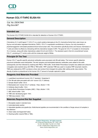
Human CCL17/TARC ELISA Kit Cat. No.:DEIA7849 Pkg.Size:96T Intended use The Human CCL17/TARC ELISA Kit is intended for detection of Human CCL17/TARC. General Description Chemokine (C-C motif) ligand 17 (CCL17) is a small cytokine belonging to the CC chemokine family that is also known as thymus and activation regulated chemokine (TARC). CCL17 is expressed constitutively in thymus, but only transiently in phytohemagglutinin stimulated peripheral blood mononuclear cells. This chemokine specifically binds and induces chemotaxis in T cells and elicits its effects by interacting with the chemokine receptor CCR4. The gene for CCL17 is located on chromosome 16, in humans, along with other chemokines called CCL22 and CX3CL1. The standard used in this kit is recombinant human CCL17, consisting of 71 amino acids with the molecular weight of 8Kda. Principle Of The Test Human CCL17 specific-specific polyclonal antibodies were precoated onto 96-well plates. The human specific detection polyclonal antibodies were biotinylated. The test samples and biotinylated detection antibodies were added to the wells subsequently and then followed by washing with PBS or TBS buffer. Avidin-Biotin-Peroxidase Complex was added and unbound conjugates were washed away with PBS or TBS buffer. HRP substrate TMB was used to visualize HRP enzymatic reaction. TMB was catalyzed by HRP to produce a blue color product that changed into yellow after adding acidic stop solution. The density of yellow is proportional to the human CCL17 amount of sample captured in plate. Reagents And Materials Provided 1. Lyophilized recombinant human CCL17 standard: 10ng/tube×2. 2. One 96-well plate precoated with anti- human CCL17 antibody. 3. Sample diluent buffer: 30 ml 4. Biotinylated anti- human CCL17 antibody: 130μl, dilution 1:100. 5. Antibody diluent buffer: 12ml. 6. Avidin-Biotin-Peroxidase Complex (ABC): 130μl, dilution 1:100. 7. ABC diluent buffer: 12ml. 8. TMB color developing agent: 10ml. 9. TMB stop solution: 10ml. Materials Required But Not Supplied 1. Microplate reader in standard size. 2. Automated plate washer. 3. Adjustable pipettes and pipette tips. Multichannel pipettes are recommended in the condition of large amount of samples in the detection. 4. Clean tubes and Eppendorf tubes. 5. Washing buffer (neutral PBS or TBS). Creative Diagnostics. All rights reserved 45-16 Ramsey Road Shirley, NY 11967, USA Tel: 631-624-4882 ·Fax:631-614-7828 E-mail: info@creative-diagnostics.com www.creative-diagnostics.com 1
Preparation of 0.01M TBS: Add 1.2g Tris, 8.5g Nacl; 450 μl of purified acetic acid or 700 μl of concentrated hydrochloric acid to 1000ml H2O and adjust pH to 7.2-7.6. Finally, adjust the total volume to 1L. Preparation of 0.01 M PBS: Add 8.5g sodium chloride, 1.4g Na2HPO4 and 0.2g NaH2PO4 to 1000ml distilled water and adjust pH to 7.2 -7.6. Finally, adjust the total volume to 1L. Storage Store at 4℃ for frequent use, at -20℃ for infrequent use. Avoid multiple freeze-thaw cycles (Shipped with wet ice.) Specimen Collection And Handling • Sample Preparation and Storage Store samples to be assayed within 24 hours at 2-8℃. For long-term storage, aliquot and freeze samples at -20℃. Avoid repeated freeze-thaw cycles. Cell culture supernate, tissue lysate or body fluids: Remove particulates by centrifugation, analyze immediately or aliquot and store at -20℃ Serum: Allow the serum to clot in a serum separator tube (about 30 min) at room temperature. Centrifuge at approximately 1000 X g for 15 min. Analyze the serum immediately or aliquot and store frozen at -20℃. Plasma: Collect plasma using EDTA as an anticoagulant. Centrifuge for 15min at 1000 x g within 30 min of collection. Analyze immediately or aliquot and store samples at -20℃. • Sample Dilution Guideline The user needs to estimate the concentration of the target protein in the sample and select a proper dilution factor so that the diluted target protein concentration falls near the middle of the linear regime in the standard curve. Dilute the sample using the provided diluent buffer. The following is a guideline for sample dilution. Several trials may be necessary in practice. The sample must be well mixed with the diluents buffer. High target protein concentration (20-200ng/ml). The working dilution is 1:100. i.e. Add 3 μl sample into 297 μl sample diluent buffer. Medium target protein concentration (2-20ng/ml). The working dilution is 1:10. i.e. Add 25 μl sample into 225 μl sample diluent buffer. Low target protein concentration (31.2-2000pg/ml). The working dilution is 1:2. i.e. Add 100 μl sample to 100 μl sample diluent buffer. Very Low target protein concentration (≤31.2pg/ml). No dilution necessary, or the working dilution is 1:2. Reagent Preparation • Reagent Preparation and Storage A. Reconstitution of the human CCL17 standard: CCL17 standard solution should be prepared no more than 2 hours prior to the experiment. Two tubes of CCL17 standard (10ng per tube) are included in each kit. Use one tube for each experiment. a. 10,000pg/ml of human CCL17 standard solution: Add 1 ml sample diluent buffer into one tube, keep the tube at room temperature for 10 min and mix thoroughly. b. 2000pg/ml of human CCL17 standard solution: Add 0.2 ml of the above 10ng/ml CCL17 standard solution into 0.8 ml sample diluent buffer and mix thoroughly. c. 1000pg/mlP31.2pg/ml of human CCL17 standard solutions: Label 6 Eppendorf tubes with 1000pg/ml, 500pg/ml, 250pg/ml, 125pg/ml, 62.5pg/ml, 31.2pg/ml, respectively. Aliquot 0.3 ml of the sample diluent buffer into each tube. Add 0.3 ml of the above 2000pg/ml CCL17 standard solution into 1st tube and mix. Transfer 0.3 ml from 1st tube to 2nd tube and mix. Transfer 0.3 ml from 2nd tube to 3rd tube and mix, and so on. Note: The standard solutions are best used within 2 hours. The 10 ng/ml standard solution may be stored at 4℃ for up to 12 Creative Diagnostics. All rights reserved 45-16 Ramsey Road Shirley, NY 11967, USA Tel: 631-624-4882 ·Fax:631-614-7828 E-mail: info@creative-diagnostics.com www.creative-diagnostics.com 2
hours, or at -20℃ for up to 48 hours. Avoid repeated freeze-thaw cycles. B. Preparation of biotinylated anti-human CCL17 antibody working solution: The solution should be prepared no more than 2 hours prior to the experiment. a. The total volume should be: 0.1ml/well x (the number of wells). (Allowing 0.1-0.2 ml more than total volume) b. Biotinylated anti-human CCL17 antibody should be diluted in 1:100 with the antibody diluent buffer and mixed thoroughly. (i.e. Add 1 μl Biotinylated anti-human CCL17 antibody to 99 μl antibody diluent buffer.) C. Preparation of Avidin-Biotin-Peroxidase Complex (ABC) working solution: The solution should be prepared no more than 1 hour prior to the experiment. a. The total volume should be: 0.1ml/well x (the number of wells). (Allowing 0.1-0.2 ml more than total volume) b. Avidin- Biotin-Peroxidase Complex (ABC) should be diluted in 1:100 with the ABC dilution buffer and mixed thoroughly. (i.e. Add 1μl ABC to 99 μl ABC diluent buffer.) Assay Steps 1. Aliquot 0.1ml per well of the 2000pg/ml, 1000pg/ml, 500pg/ml, 250pg/ml, 125pg/ml, 62.5pg/ml, 31.2pg/ml human CCL17 standard solutions into the precoated 96-well plate. Add 0.1ml of the sample diluent buffer into the control well (Zero well). Add 0.1ml of each properly diluted sample of human sera, plasma, body fluids, tissue lysates or cell culture supernatants to each empty well. We recommend that each human CCL17 standard solution and each sample is measured in duplicate. 2. Seal the plate with the cover and incubate at 37℃ for 90 min. 3. Remove the cover, discard plate content, and blot the plate onto paper towels or other absorbent material. Do NOT let the wells completely dry at any time. 4. Add 0.1ml of biotinylated anti-human CCL17 antibody working solution into each well and incubate the plate at 37℃ for 60 min. 5. Wash plate 3 times with 0.01M TBS or 0.01M PBS, and each time let washing buffer stay in the wells for 1 min. Discard the washing buffer and blot the plate onto paper towels or other absorbent material. Plate Washing Method: Discard the solution in the plate without touching the side walls. Blot the plate onto paper towels or other absorbent material. Soak each well with at least 0.3 ml PBS or TBS buffer for 1~2 minutes. Repeat this process two additional times for a total of THREE washes. Note: For automated washing, aspirate all wells and wash THREE times with PBS or TBS buffer, overfilling wells with PBS or TBS buffer. Blot the plate onto paper towels or other absorbent material.″ 6. Add 0.1ml of prepared ABC working solution into each well and incubate the plate at 37℃ for 30 min. 7. Wash plate 5 times with 0.01M TBS or 0.01M PBS, and each time let washing buffer stay in the wells for 1-2 min. Discard the washing buffer and blot the plate onto paper towels or other absorbent material. (See Step 5 for plate washing method). 8. Add 90 μl of prepared TMB color developing agent into each well and incubate plate at 37℃ in dark for 15-20 min (Note: For reference only, the optimal incubation time should be determined by end user. And the shades of blue can be seen in the wells with the four most concentrated human CCL17 standard solutions; the other wells show no obvious color). 9. Add 0.1ml of prepared TMB stop solution into each well. The color changes into yellow immediately. 10. Read the O.D. absorbance at 450nm in a microplate reader within 30 min after adding the stop solution. For calculation, (the relative O.D.450) = (the O.D.450 of each well) – (the O.D.450 of Zero well). The standard curve can be plotted as the relative O.D.450 of each standard solution (Y) vs. the respective concentration of the standard solution (X). The human CCL17 concentration of the samples can be interpolated from the standard curve. Note: if the samples measured were diluted, multiply the dilution factor to the concentrations from interpolation to obtain the concentration before dilution. Typical Standard Curve Typical Data Obtained from Human CCL17 Table 1. Creative Diagnostics. All rights reserved 45-16 Ramsey Road Shirley, NY 11967, USA Tel: 631-624-4882 ·Fax:631-614-7828 E-mail: info@creative-diagnostics.com www.creative-diagnostics.com 3
Figure 1. Detection Range 31.2pg/ml-2000pg/ml Sensitivity < 1pg/ml Specificity No detectable cross-reactivity with any other cytokine. Precautions 1. Before using Kit, spin tubes and bring down all components to bottom of tube. 2. Duplicate well assay was recommended for both standard and sample testing. 3. Don’t let 96-well plate dry, dry plate will inactivate active components on plate. 4. In order to avoid marginal effect of plate incubation due to temperature difference ( reaction may be stronger in the marginal wells), it is suggested that the diluted ABC and TMB solution will be pre-warmed in 37℃ for 30 min before using. Creative Diagnostics. All rights reserved 45-16 Ramsey Road Shirley, NY 11967, USA Tel: 631-624-4882 ·Fax:631-614-7828 E-mail: info@creative-diagnostics.com www.creative-diagnostics.com 4
Analyte Gene Information Gene Name CCL17 chemokine (C-C motif) ligand 17 [ Homo sapiens ] CCL17 CCL17; chemokine (C-C motif) ligand 17; SCYA17, small inducible cytokine subfamily A (Cys Cys), member 17; C-C motif chemokine 17; ABCD 2; TARC; CC chemokine TARC; T cell-directed CC chemokine; small-inducible cytokine A17; thymus and activation-regulated chemokine; small inducible cytokine subfamily A (Cys-Cys), member 17; ABCD-2; SCYA17; A-152E5.3; MGC138271; MGC138273; Official Symbol Synonyms GeneID 6361 mRNA Refseq NM_002987 Protein Refseq NP_002978 MIM 601520 Q92583 UniProt ID Chromosome Location 16q13 Chemokine receptors bind chemokines, organism-specific biosystem; Chemokine signaling pathway, organism-specific biosystem; Chemokine signaling pathway, conserved biosystem; Class A/1 (Rhodopsin-like receptors), organism-specific biosystem; Cytokine-cytokine receptor interaction, organism-specific biosystem; Cytokine-cytokine receptor interaction, conserved biosystem; GPCR ligand binding, organism-specific biosystem; chemokine activity; receptor binding; Pathway Function REFERENCES 1. Imai T, Yoshida T, Baba M, Nishimura M, Kakizaki M, Yoshie O (1996). "Molecular cloning of a novel T cell-directed CC chemokine expressed in thymus by signal sequence trap using Epstein-Barr virus vector". J. Biol. Chem. 271 (35): 21514–21. 2. Imai T, Baba M, Nishimura M, Kakizaki M, Takagi S, Yoshie O (1997). "The T cell-directed CC chemokine TARC is a highly specific biological ligand for CC chemokine receptor 4". J. Biol. Chem. 272 (23): 15036–42. Creative Diagnostics. All rights reserved 45-16 Ramsey Road Shirley, NY 11967, USA Tel: 631-624-4882 ·Fax:631-614-7828 E-mail: info@creative-diagnostics.com www.creative-diagnostics.com 5