Alteration in fluids & electrolytes
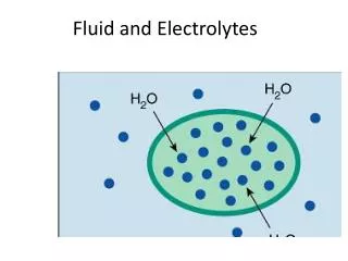
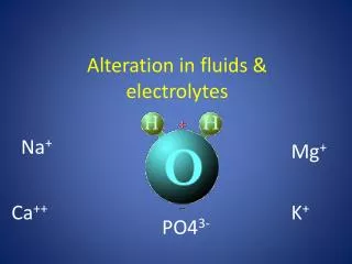
Alteration in fluids & electrolytes. Na +. Mg +. K +. Ca ++. PO4 3-. Body fluid compartments. Total body water (TBW). 75. 25. Electrolytes. Intracellular fluid has higher concentration of potassium, calcium, phosphates and magnesium .

Alteration in fluids & electrolytes
E N D
Presentation Transcript
Alteration in fluids & electrolytes Na+ Mg+ K+ Ca++ PO43-
Body fluid compartments Total body water (TBW) 75 25
Electrolytes • Intracellular fluid has higher concentration of potassium, calcium, phosphates and magnesium. • Extracellular fluid has higher concentration of sodium, chloride and bicarbonate.
The movement of body fluids between the ICF and • ECF compartments occurs at the cell membrane and depends • on regulation of ECF water and sodium. • Regulation of fluid volume, solute concentration and distribution between the 2 compartments depends on water and sodium balance
Homeostasis maintained by: Mainly by Kidney function Ion transport Water movement
Water Movement • Cell membranes are semipermeable allowing water to pass through • Osmosis- major way fluids transported Water shifts from low solute concentration to high solute concentration to reach homeostasis (balance).
OSMOSIS • Movement of fluid across membrane from a lower solute concentration to a higher solute concentration • Passive process
Osmolarity • Concentration of particles in solution • The greater the concentration (Osmolarity) of a solution, the greater the pulling force (Osmotic pressure) • Normal blood osmolarity = 280-295 mOSM/kg • A solution that has HIGH osmolarity is one that is > blood osmolarity = HYPERTONIC solution • A solution that has LOW osmolarity is one that is < blood osmolarity = HYPOTONIC solution • A solution that has equal osmolarity as blood = ISOTONIC solution
Solutes – Dissolved Particles Electrolytes – charged particles Cations – positively charged ions Na+, K+ , Ca++, H+ Anions – negatively charged ions Cl-, HCO3- , PO43- Non-electrolytes - Uncharged Proteins (i.e. albumin), urea, glucose, O2, CO2 10
Solute Movement - Diffusion • Movement of solutes from high concentration to low concentration • It is a PASSIVE movement DOWN the concentration gradiant. (requires no energy)
DIFFUSION • Solutes shift from an area of higher concentration to an area of lower concentration • Passive process
Solute Movement –other mechanisms Active transport- requires energy (ATP) to move from low concentration to high concentrationExample: Na / K pump • May be enhanced by carrier molecules with binding sites on cell membraneExample: Glucose (Insulin promotes the insertion of binding sites for Glucose on cell membranes).
Regulation of body water The default is get rid of it The control processes include: Release of ADH (antidiuretic hormone) Thirst
Regulation of body water • Any of the following: • Decreased amount of water in body • Increased amount of Na+ in the body • Increased blood osmolality • Decreased circulating blood volume • Results in: • Stimulation of osmoreceptors in hypothalamus • Release of ADH from the posterior pituitary • Increased thirst • And thus: water consumption and conservation
Regulation of Sodium Sodium is largely regulated by the kidney under the influence of the sympathetic nervous system and the rennin-angiotensin-aldosterone mechanism.
Alterations of sodium and water balance can be divided • into two main categories: • isotonic disorders: contraction or expansion of ECF volume (confined to the ECF compartment) • hypotonic dilution (dilutionalhyponatremia) or hypertonic concentration (hypernatremia) of extracellular sodium brought about by changes in extracellular water
Volume Abnormalities Isotonic fluid loss • ↓ECF volume, • it causesthirst, dry skin and mucous membranes, ↓ urine output decreased vascular volume and circulatory function, and increased urine specific gravity: hypovolemic shock
Volume Abnormalities Isotonic fluid gain • ↑ECF volume, ↑ B.P, increased vascular volume andOedema
electrolyte balance • Electrolytes help to regulate myocardial and neurological function, fluid balance, acid base balance • The most common cause of electrolyte disturbances is renal failure. • The most serious electrolyte disturbances involve abnormalities in the levels of sodium, potassium, and/or calcium. • Chronic laxative abuse or severe diarrhea or vomiting can lead to electrolyte disturbances along with dehydration.
Electrolyte balance • Na + (Sodium) • Predominant extracellular cation • Pairs with Cl- , HCO3- to neutralize charge • Most important ion in water balance • Important in nerve and muscle function • Reabsorption in renal tubule regulated by: • Sympathatic nervous system • Renin/angiotensin- Aldosterone
Hyponatraemia • Decrease in Plasma Na+ • Due to ↓Na + or ↑water • Water moves from ECF → ICF → Cells swell • Aetiology: • Excessive sodium loss • Exercise or heat induced sweating • Gastrointestinal loss (vomiting, diarrhea) • Excessive water intake in relation to output • Excessive administration of sodium-free parenteral solutions. • Kidney disorders that impair water elimination • Increased ADH levels (stress, pain, use of medications that increase ADH)
Clinical manifestations • Due to movement of extracellular fluids and water into brain cells and neuromuscular tissue causing: • Muscle cramps, weakness, headache, confusion, convulsion, depression, fatigue and coma
Hypernatremia • Plasma Na+ > 145 mEq / L • Due to ↑ Na + or ↓ water • Water moves from ICF → ECF Cells dehydrate Due to: • Excess Na intake : administration of hypertonic IV solution) • Excess Na retention (oversecretion of aldosterone Conn’s syndrome, Cushing’s syndrome) • Loss of pure water • diuretics • Diabetes insipidus – polyuria • Insufficient intake of water (hypodipsia)
Clinical manifestations are due to shrinking of body cells, including those of the CNS • Fatigue, weakness. With more severe elevations of the sodium level, seizures and coma may occur.
Electrolyte balance • K + (Potassium) Major intracellular cation The distribution between the intracellular and the extracellular compartments regulates: • electrical membrane potentials controlling the excitability of nerve and muscle cells • contractility of skeletal and • cardiac muscle and smooth muscle. Regulation in kidney through: Aldosterone Transcellular shift
Hypokalemia • is a decrease in serum potassium level • Aetiology: • Inadequate intake, excessive gastrointestinal and skin loss • Excessive renal loss • Diuretics • Increased mineralocorticoid levels (Conn’s syndrome, treatment with corticosteroid therapy) • Low level of magnesium • Extracellular to intracellular shift: administration of insulin • Clinical manifestations: • A low potassium level can make muscles feel weak, cramp, twitch, or even become paralyzed • Change in the electrocardiogram and abnormal heart rhythms may develop.
Hyperkalemia • is the increase in serum potassium level • Aetiology: • Release from intracellular compartment • Excessive intake (rapid infusion of K containing parenteral fluid) • Inadequate elimination by the kidney such as in renal failure, hypoaldosteronism, treatment with Angiotensin-converting enzyme inhibitor • Clinical manifestations: • When hyperkalemia becomes more severe, it can cause abnormal heart rhythms. • Change in the electrocardiogram. Risk of cardiac arrest with severe excess.
Calcium (Ca++) Calcium is essential for the formation of bones and teeth, muscle contraction, blood clotting , neuromuscular transmission and function of many enzymes. Calcium balance • About 99% of the body's calcium is stored in bones, 0.7% is located inside cells and 0.2 to 0.3% is found in ECF. • Calcium in the ECF is either free (ionized), complexed or protein bound. • Only the ionized form play an essential role in neuromuscular transmission • The level of calcium in blood is regulated primarily by two hormones: parathyroid hormone and calcitonin. .
Parathyroidhormone is produced by the four parathyroid glands, located around the thyroid gland in the neck. • When the calcium level in blood decrease, more parathyroid hormone is produced and when calcium level increase, parathyroid hormone decrease • Effect of parathyroid hormone: • Stimulates bones to release calcium into blood • Causes the kidneys to excrete less calcium in urine • Stimulates the digestive tract to absorb more calcium • Causes the kidneys to activate vitamin D, which enables the digestive tract to absorb more calcium • Calcitonin is produced by cells of the thyroid gland. It lowers the calcium level in blood by slowing the breakdown of bone.
Calcium balance • Most in ECF • Regulated by: • Parathyroid hormone • ↑Blood Ca++ by stimulating osteoclasts • ↑GI absorption and renal retention • Calcitonin from the thyroid gland • Promotes bone formation • ↑ renal excretion
Hypocalcemia • In hypocalcemia, the calcium level in blood is too low. • Aetiology: • Impaired ability to mobilize calcium from bone • A low level of parathyroid hormone (hypoparathyroidism), • A low level of magnesium (hypomagnesemia) • Decreased intake or absorption • Inadequate consumption of calcium • Vitamin D deficiency • Medications that impair activation of vitamin D: rifampin (an antibiotic), anticonvulsants (such as phenytoin) • Abnormal renal losses • Kidney dysfunction and hyperphosphatemia • Increased sequestration • Pancreatitis
Clinical manifestations • Increase neuromuscular excitability • Hypocalcemia can affect the brain and cause neurologic or psychologic symptoms, such as confusion, memory loss, depression, and hallucinations. These symptoms disappear if the calcium level is restored. • An extremely low calcium level may cause tingling (often in the lips, tongue, fingers, and feet), muscle aches, spasms of the muscles in the throat (leading to difficulty breathing), seizures, and abnormal heart rhythms.
Hypercalcemia • In hypercalcemia, the calcium level in blood is too high. • Aetiology: • Increased intestinal absorption • Excessive vit D • Excessive calcium in diet • Milk alkali syndrome (patient with peptic ulcers drinking milk and taking calcium-containing antacids for relief). • Increased bone resorption • Increased level of parathyroid hormone, malignacy, prolonged immobilization • Decreased elimination (thiazide diuretics)
Clinical manifestations: • Exposure of kidney to increased concentration of calcium (signs of kidney stones) • Gastrointestinal manifestations (constipation, nausea, vomiting, and loss of appetite). • Decrease neuromuscular excitability (muscle weakness and atrophy)
Phosphate Phosphate balance • Phosphate is an intracellular anion • 85% of the phosphorus is contained in bone. It is necessary for the formation of bone and teeth. • the remaining in organic compounds such as nucleic acids and high energy compounds (e.g. ATP). • Many of the manifestations of hypophosphatemia are due to ATP depletion. • Serum levels of calcium and phosphate are inversely proportional to prevent the damaging deposition of calcium phosphate in soft tissues. • Many of the manifestations of hyperphosphatemia reflect a decrease in serum calcium levels
HYPOPHOSPHATEMIA In hypophosphatemia, the level of phosphate in blood is too low. • Aetiology: • Decrease intestinal absorption: antacids (aluminium and calcium),severe diarrhea, lack of vitamin D • Increased renal elimination: Hyperparathyroidism, impaired kidney function • Malnutrition and intracellular shift • Clinical manifestations: • Neural dysfunction (confusion and coma), • Disturbed musculoskeletal function (muscle weakness, bone pain) • Hematological disorders (anemia, platelet dysfunction, impaired white blood cell function).
HYPERPHSPHATEMIA • In hyperphosphatemia, the level of phosphate in blood is too high. • Hyperphosphatemia is rare except in people with severe kidney dysfunction. • Aetiology: • Impaired elimination (kidney failure, hypoparathyroidism) • Acute phosphate overloads (enemas containing phosphate, intravenous phosphate supplementation) • Clinical manifestations: • Hyperphosphatemia is associated with decreased in serum calcium. • Many of the sign and symptoms of hyperphosphatemia are related to hypocalcemia.
Magnesium (Mg+) • Most of the body magnesium is located within cells • it functions in regulation of enzyme activity, and calcium transport • Magnesium is necessary for parathyroid hormone function and hypomagnesemia is a common cause of hypocalcemia • Thus magnesium is necessary for the formation of bone and teeth • There is interdependency between intracellular concentrations of magnesium and potassium such that a decrease in one is accompanied by a decrease in the other. • Magnesium deficiency contributes to cardiac dysrhysmiasisthat occur with hypokalemia.
HYPOMAGNESEMIA • In hypomagnesemia, the level of magnesium in blood is too low. • hypomagnesemia impairs PTH release and the action of PTH • It leads to reduction in intracellular potassium and impairs the ability of the kidney to conserve potassium • Aetiology: • Impaired intake or absorption: • malnutrition or starvation, • malabsorption, • Parenteralhyperalimentation with inadequate amounts of magnesium • Increased loss • Diuretics • Hyperparathyroidism, hyperaldosteronism • Clinical manifestations: The signs and symptoms of hypomagnesemia are similar to hypocalcemia
HYPERMAGNESEMIA • In hypermagnesemia, the level of magnesium in blood is too high. • Hypermagnesemia usually develops only when people with kidney failure are given magnesium salts (mineral supplements) or the administration of drugs that contain magnesium (such as some antacids or laxatives). • It causes neuromuscular dysfunction, and muscle weakness. • Magnesium decreases acetylcholine release at the myoneural junction and may cause neuromuscular blockade and respiratory paralysis. • Severe cases can lead to cardiac arrest.