Download

1 / 27
270 likes | 350 Vues
Explore the external and general features of the cerebellum, including its functional subdivisions: archicerebellum, paleocerebellum, and neocerebellum. Learn how each part influences equilibrium, muscle tone, posture, and movement coordination.

E N D
General Features of Cerebellum : • The cerebellum consists of a midline vermis and 2-lateral hemispheres. • Anatomically , it is divided into anterior, posterior& flocculo-nodular lobes. • It controls equilibrium, it influences posture & muscle tone and coordinates the movements • Its surface is high convoluted, forming folds or folia, being oriented transversely • It lies behind Pons & M.O. , separated from them by the cavity of 4th ventricle. • It is connected to brain stem (medulla, pons& midbrain) by inferior,middle & superior cerebellarpedunclesrespectively. Lateral aspect of brain stem & cerebellum , showing cerebellar peduncles.
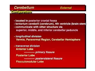
External Features of Cerebellum : • It has anterior notch ,which is wider and lodging the back of pons& medulla. It is separated from them by cavity of 4thventricle • It has also posterior notch occupied by falx cerebelli, which separates the 2 cerebellar H. • Inferior surface : rounded oneach side and presents : a deep groove (vallecula) between the 2-cerebellar hemispheres,which is occupied by the inferior vermis.-Tonsil isasmall part of cerebellar hemisphere that lies lateral to inferior vermis. Superior surface Inferior surface
Superior surface :lies beneath tentorium cerebelli and has a raised superior vermis + a large cerebellar hemisphere on each side + primary & horizontal fissures.1-Primary fissure V-shaped,well defined fissure, lies on superior surface and separates the small anterior lobe from the larger middle lobe (or posterior lobe).2- Horizontal fissurelies along the sides ofcerebellum, extending from anterior notch to posterior notch, separates the superior from the inferior surfaces. External Features of Cerebellum : 3- Secondary (posterolateral) fissurelies on inferior surface and separates flocculo-nodular lobe from the ramainder of cerebellum.
Functional subdivision of cerebellum : 1- Archi-cerebellum = posterior lobe (Vestibular part) : _ It is formed of the flocculo-nodular lobe + associated fastigialnuclei, lying on inf. Surface in front of postero-lateral fissure._Embryologically, it is the oldest part of cerebellum._It receives afferent Fs. From vestibular apparatus of internal ear Via vestibulo-cerebellar tracts._It is concerned with equlibrium. • Schematic drawing of cerebellum showing the relationshipsbetween the anatomical & functional divisions of cerebellum. • Green =archi-cerebellum, blue=paleo-cerebellum. Pink= neo-cerebellum
I- Archicerebellum • It is concerned with equilibrium. • It represents flocculo-nodular lobe. • It has connections with vestibular &reticular nuclei of brain stem through the inferior cerebellar peduncle. • Afferent vestibular Fs. Pass from vestibular nuclei in pons & medulla to the cortex of ipsilateral flocculo-nodular lobe. • Efferent cortical (purkinje cell) Fs. Project to fastigial nucleus, which projects to vestibular nuclei & reticular formation. • It affects the L.M.system bilaterally via descending vestibulo-spinal & reticulo-spinal tracts. Connections of archicerebellum
2- Paleo-cerebellum= (spinal part) : -_it is formed of midline vermis + surrounding paravermis + globose & emboliform nuclei._It receives afferentproprio-ceptive impulses from Ms.& tendons Via spino-cerebellar tracts (dorsal & ventral) mainly.-it sends efferents to red nucleus of midbrain. -it is concerned withmuscle tone • Schematic drawing of cerebellum showing the relationshipsbetween the anatomical & functional divisions of cerebellum. • Green =archi-cerebellum, blue=paleo-cerebellum. Pink= neo-cerebellum
2-Paleo-cerebellum • It is concerned with muscle tone& posture. • Afferents spinal Fs. consist of dorsal & ventral spino-cerebellar tract from muscle, joint & cutaneous receptors to enter the cortex of ipsilateralvermis & para vermis Viainferior & superior cerebellar peduncles . • Efferents cortical fibres pass to globose & emboliform nuclei, then Via sup. C. peduncle to contra-lateral rednucleus of midbrain to give rise descending rubro-spinal tract. Connections of Paleo-cerebellum.
3- Neo-cerebellum= (cerebral part) : _It is the remaining largest part of cerebellum. _It includes the most 2-cerebellarhemispheres+ dendate nuclei._It receives afferent impulses from the cerebral cortex+pons Via cerebro-ponto- cerebellar pathway. -it sends efferents to V.L.nucleus of thalamus. -it controls voluntary movements(muscle coordination). • Schematic drawing of cerebellum showing the relationshipsbetween the anatomical & functional divisions of cerebellum. • Green =archi-cerebellum, blue=paleo-cerebellum. Pink= neo-cerebellum
3- Neo-cerebellum • It is concerned withmuscular coordination. • It receives afferentsfrom cerebral cortex involved in planning of movement- to pontine nuclei ,cross to opposite side Via middle Cerebellarpeduncle to end in lateral parts of cerebellum (cerebro-ponto-cerebellar tract). • Neo-cerebellar efferents project to dendatenucleus,which in turn projects to contra-lateral red nucleus & ventral lateral nucleusof thalamus ,then to motor cortex of frontal lobe, giving rise descending cortico-spinal & cortico-bulbar pathways.Efferents of dentate nucleus form a majorpart of superior C. peduncle. Connections of Neo-cerebellum.
Cerebellar Lesions • Are usually vascular, may be traumatic or tumour. • Manifestations of unilateral cerebellar lesions :1-ipsilateral incoordination of (U.L) arm = intention tremors : it is a terminal tremors at the end of movement as in touching nose or button the shirt. 2-Or ipsilateral cerebellar ataxia affects (L.L.) leg, causing wide-based unsteady gait. • Manifestations of bilateral cerebellar lesions (caused by alcoholic intoxication, hypothyrodism, cerebellar degeneration & multiple sclerosis) :1-dysarthria : slowness & slurring of speech. 2-Incoordination of both arms.=intention tremors.3-Cerebellar ataxia : intermittent jerky movements or staggering , wide-based, unsteady gait. 4-Nystagmus : is a very common feature of multiple sclerosis. It is due to impairment coordination of eye movements /so, incoordination of eye movements occurs and eyes exhibit a to-and-fro motion. • Combination of nystagmus+ dysarthria + intension tremors constitutes Chacot’triad, which is highly diagnostic of the disease.
Internal Structure of cerebellum : • It consists of an outerlayer ofgrey matter(cerebellar cortex) , & inner layer ofwhite matter containing 4-pairs ofcerebellar nuclei :above roof of 4th V. from medial to lateral :1-Fastigial nucleus. 2-Globose nucleus. 3-Emboliform nucleus. 4-Dendate nucleus.(theonly one that can be seen clearly with the nakedeye). • Sagittal section of cerebellum. • T.S.of cerebellum & brain at level of 4th V. to show cerebellar nuclei.
Cerebellar cortex • It is highly convoluted, forming numerous transverselyoriented folia. • It contains nerve cells,dendrites and synaptic connections of cellular neurones. • The cellular organization of the cortex consists of 3-layers : 1-Outer molecular layer. 2-Intermediate, purkinje celllayer. 3-Inner granular layer, which is dominated by granule cell. T.S of cerebellar folia showing layers of cerebellar cortex. Afferent & Efferent connecltions and their relationships to principal cells of cerebellar cortex.
Cerebellar cortex • Molecular layer : contains 1-Cells : molecular cells (stellatecells) & basket cells. 2-Nerve Fibres :a- dendrites of Purkinje cells (arborisations). B-axons of granule cells. ( bifurcateto produce 2-parallel fibres , oriented along long axis of folium). C-ending of climbing fibers. • Purkinje cell layer :it isformed of one layer (unicellular) of large flask-shaped purkinje cells. Their arborisations are at right angles to long axis to folium. • Granular layer :it is formedof small granule cells & ending of mossy fibres.
M • There are 3-types of Nerve Fibres in white Matter :1-Axons of purkinje cells : the only axons to leave cerebellar cortex toend in deep cerebellarnuclei specially dendate nucleus. 2-Mossy Fibres :end in thegranularlayer. 3-Climbing Fibres : end in the molecular layer.
Afferent Fibres to cerebellum : • Mostly end in cerebellarcortex, excitatory to cortical neurones, as mossy or climbing Fs. passing through the cerebellar peduncles. • The following are Afferent fibres:1-dorsal & ventral spino-cerebellar tract. (passing via I.C.P & S.C.P) 2-vestibulo-cerebellar Fs. (via I.C.P) 3-olivo-cerebellar Fs. (via I.C.P)/ (extrapyramidal fibres),(end as climbing or mossy fibres) 4-ponto-cerebellar Fs. (via M.C.P). (In M.O)
Efferent Fibres of the cerebellum : • It sends the following fibres : 1-Cerebello-vestibular Fs. to vestibular nuclei of pons & M.O. 2-Cerebello-olivary Fs. To M.O. 3-Dendato-rubro-thalamic tract To red nucleus of midbrain & ventro-lateral nucleus of the thalamus and finally to motor cortex of frontal lobe to coordinate movement via cortico-spinal & corticobulbar tracts. M
The Fourth Ventricle • It is a cavity of hindbrain. • Position : lies between pons & M.O. anteriorly and the cerebellum posteriorly. • It is a diamond-shaped space which is lined by ependyma. • Its superior angle is continuous with cerebral aqueduct of midbrain. • inferior angle is continuous with centeral canal ofclosed M.O. • Its lateral angles extend laterally toform a lateral recess on each side to open into subarachnoid space.
The Boundaries of 4th Ventricle • Superiolateral boundary : -it is formed by superior cerebellarpeduncle on each side. • Inferiolateral boundary : -it is formed by inferior cerebellarpeduncle + gracile & cuneatetubercles on each side.
The Roof of 4th Ventricle -it is a tent-shaped when seen laterally, and diamond-shaped when seen behind. -it is formed of superior &inferior medullary vela, which are thin sheets of white matter /consists of : ependyma covered by pia mater. -Sup.medullary velum connects the 2 sup.cerebellar peduncles. -Inf.medullary velum connects the 2 inf.cerebellar peduncles. -Inferior vermis of cerebellum : lies in the middle of roof of 4th ventricle.
The Roof of 4th Ventricle • The lower part of roof is invaginated by tela choroidea of 4th ventricle. • The tela choroidea is a double layer of pia mater which encloses the choroid plexus of 4th ventricle. • The choroid plexus is a vascularcapillary tuft covered by ependymal cells and secretes C.S.F. into the lumen of 4th ventricle.
The Openings of 4th Ventricle • The roof contains 3 aperatures which transmit C.S.F. from ventricular lumen to subarachnoid space. • Median aperature (foramen of Magendie) : lies in the median plane at lower end of inferior medullary velum, and opens into subarachnoid space at cistrna magna at cerebello-medullary angle • 2 lateral openings (foramina of Luschka) : each one lies at the lateral end oflateral recess to open intosubarachnoid space atcerebello-pontineangle. choroid plexus partly protrudes out through each lateral aperture.
The Floor of 4th Ventricle • It is called rhomboid fossa. • It is diamond-shaped and is divided into right & left halves by the median sulcus. • It is crossed in the middle by transvere Fs. (ponto-cerebellar Fs.)called medullary stria, which divide floor of 4th ventricle into upper (pontine) &lower (medullary) part. A diagram to show the floor & lateral boundaries of 4th ventricle.
The Floor of 4th Ventricle • Upper pontine part : presents on each side of median sulcus. 1-Medial eminence : a rounded elevation produced by the abducent nucleus.2-Facial colliculus : an elevation on the top of lower part of medial eminence. It is produced by the fibres of facial nerve which surround abducent nucleus. 3-Superior fovea : a groove lateral to facial colliculus. 4-Vestibular area : lateral to superior fovea. It overlies superior, medial & lateral vestibular nuclei. A diagram to show the floor & lateral boundaries of 4th ventricle.
The Floor of 4th Ventricle • Lower medullary part : presents on each side of the median sulcus. 1-Inferior fovea : inverted V-shaped groove. 2-Hypoglossal area : medial to inferior fovea. It overlies hypo-glossal nucleus.3-Vagal area (triangle) : between limbs of inferior fovea.It overlies dorsal nucleus of vagus.4-Vestibular area : lateral to inferior fovea. It overlies inferiorvestibular nucleus. A diagram to show the floor & lateral boundaries of 4th ventricle.