Download
1 / 73
990 likes | 2.06k Vues
Cardiovascular System: The Heart Chapter 19 – Lecture Notes. to accompany Anatomy and Physiology: From Science to Life textbook by Gail Jenkins, Christopher Kemnitz, Gerard Tortora. Chapter Overview. 19.1 Location & Anatomy 19.2 Heart Chambers 19.3 Heart Valves 19.4 Heart Functions

E N D
Cardiovascular System: The HeartChapter 19 – Lecture Notes to accompany Anatomy and Physiology: From Science to Life textbook by Gail Jenkins, Christopher Kemnitz, Gerard Tortora
Chapter Overview 19.1 Location & Anatomy 19.2 Heart Chambers 19.3 Heart Valves 19.4 Heart Functions 19.5 Cardiac Conduction System 19.6 ECG 19.7 Cardiac Cycle 19.8 Cardiac Output
Essential Terms cardiology • study of the cardiovascular system apex of heart • bottom point of heart formed by ventricles base of heart • formed by atria, broad superior surface ventricles • muscular chambers of the heart that eject blood atria • collecting chambers of the heart that contract to fill the ventricles
Introduction • heart beats about 100,000 times every day or about 35 million beats per year • left side pumps to systemic circuit • right side pumps to pulmonary circuit
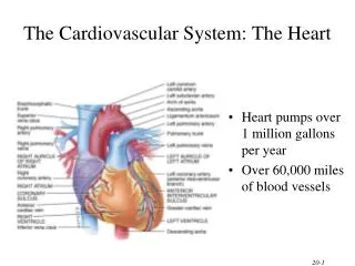
Heart Anatomy • size ~ 12cm long, 9cm wide at broadest part (5” x 3.5”) • average mass 250g (8 oz) in females • average mass 300g (10 oz) in males • rests on diaphragm • near midline of thoracic cavity • 2/3 of to left of midline • in mediastinum • between lungs • base is directed posteriorly and to right • apex
PERICARDIUM • fibrous pericardium • dense, irregular connective tissue • functions • protect and anchor serous pericardium 2. parietal pericardium • fused to the fibrous pericardium 3. visceral pericardium • also called epicardium pericardial cavity of the serous pericardium is filled with pericardial fluid
Heart Wall • epicardium • visceral layer of serous pericardium • myocardium • cardiac muscle • involuntary • branched cells • intercalated discs • gap junctions • Desmosomes • endocardium • continuous through out cardiovascular system
Right Atrium (RA) • receives blood from • superior vena cava • inferior vena cava • coronary sinus • posterior wall is smooth • anterior wall is rough with pectinate muscles that extend into auricle • divided from left atrium by thin partition called interatrial septum • oval depression in septum called fossa ovalis • remnant of foramen ovale • blood leaves RA through tricuspid valve
Right Ventricle (RV) • receives blood from right atrium • forms most of the anterior surface of heart • contains trabeaculae carneae • raised bundles of cardiac muscle • cusps of tricuspid valve connected to chordae tendineae • chordae tendineae connected to cone-shaped trabeaculae carneae called papillary muscles • divided from left ventricle by interventricular septum • blood ejected to pulmonary valve to pulmonary trunk en route to lungs for gas exchange
Left Atrium (LA) • receives blood from lungs • through 4 pulmonary veins • structurally similar to right atrium • blood passes to left ventricle through the bicuspid (mitral) valve
Left Ventricle (LV) • receives blood from LA • through bicuspid valve • internal structures similar to RV • trabeaculae carneae • chordae tendineae • papillary muscles • interventricular septum • blood ejected into aorta • some aortic blood travels to coronary arteries • remainder passes to arch of aorta • fetal life blood passes from pulmonary trunk to aorta (bypassing lungs) through ductus arteriosus (closes shortly after birth)
Myocardium • atrial walls are thinnest • right ventricle thinner than left ventricle • pumps blood shorter distance • left ventricle walls are thickest • right and left ventricles pump same volume of blood with each beat
Valves Of The Heart Ensure one way flow through the heart • Atrioventricular Valves • between the atria & the ventricles • right side - tricuspid value • left - bicuspid or mitral valve • chordae tendineae to papillary muscles • Semilunar Valves • at the beginning of the arteries that leave the heart • 3 cusps per valve • pulmonary semilunar valve • aortic semilunar valve
Systemic and Pulmonary Circulation • systemic circulation • left side of the heart • receives from lungs • pumps to aorta & body tissues • oxygenated blood • pulmonary circulation • right side of the heart • receives blood from the body tissues (veins) • pumps to pulmonary trunk & lungs • deoxygenated blood
Coronary Circulation • functional blood supply of the heart • arteries arise from base of aorta and encircle heart in atrioventricular groove • Left coronary artery • runs toward left side of heart • divides into anterior interventricular artery • supplies blood to interventricular septum and anterior walls of both ventricles • Right coronary artery • runs toward right side of heart • divides into marginal artery and posterior interventriculary artery • Marginal artery serves lateral myocardium of right side • Other serves heart apex and posterior ventricular walls
Coronary Veins • AKA coronary sinus • great cardiac vein (anterior) • middle cardiac vein (posterior) • small cardiac vein • anterior cardiac veins
Anatomy of Cardiac Conduction System • excitation begins at SA node (100/min_ • arrives at AV node located in interatrial septum, is slowed down (75/min) • AP flows to AV bundle • then enters right and left bundle branches traveling upward • final AP arrives at Purkinje fibers contracting ventricular myocardium from apex up ejecting blood through semilunar valves
CONDUCTION SYSTEM Sequence of excitation • sinoatrial (SA) node - spreads to both atria • 90 - 100 action potentials per minute • atrioventricular (AV) node • 40 -50 action potentials per minute • atrioventricular (AV) bundle (bundle of His) • 20-40 action potentials per minute • right & left bundle branches • in the interventricular septum • Purkinje fibers • conduction myofibers
Electrocardiography • recording of AP transmission through the cardiac conduction system • electrodes placed on body surface • arms and legs and six positions on chest • graphed as series of up and down waves produced during each heartbeat • instrument called electrocardiograph • produces 12 different tracings