Download
1 / 43
430 likes | 599 Vues
The Neuromuscular Hip. w mccormick june 2010. neuromuscular disorders hip development diagnosis focus on the hip history exam management nonoperative operative complications summary. Neuropathic Upper Motor Neuron CP Spinocerebellar Friedreich’s Ataxia

E N D
The Neuromuscular Hip w mccormick june 2010
neuromuscular disorders hip development diagnosis focus on the hip history exam management nonoperative operative complications summary
Neuropathic Upper Motor Neuron CP Spinocerebellar Friedreich’s Ataxia Syringomyelia Spinal Cord Tumour Trauma Myelomeningocele Lower Motor Neuron HMSN (Charcot Marie Tooth) Poliomyelitis Viral Myelitides Traumatic Spinal Muscular Atrophy Dysautonomia (Riley-Day syndrome) Myopathic Arthrogryposis Muscular Dystrophy DMD Limb-girdle Fascioscapulohumeral Fiber-type disproportion Congenital hypotonia Myotonia Dystrophica neuromuscular disorders
the problem concentric joint + balanced forces = stable hip DDH = non concentric joint starts wonky development mostly ok NM = unbalanced forces starts normal develops into wrong shape
diagnosis • spasticity/floppiness • which limbs/muscles • static vs. progressive • physical • posturing, enlarged calves, gower sign • neuro exam
testing • EMG • fibrillation with neuropathy • muscle biopsy • myopathy: variable fiber size, degeneration • neuropathy: atrophy • MR spine • for myelomengocele, syrinx
the hip - history • age • development • sypmtoms • previous treatments • MHx
the hip - exam • look • sitting/gait • check spine • movement quality • feel • tone
move • ROM • hips • abd • extension • knees • ankles
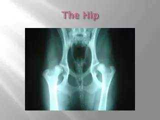
the hip - imaging • supine AP pelvis in neutral add/abd • MI • AI • femoral changes • acetabular changes • brain/spine for workup prn
treatment - nonoperative • PT for ROM monitor hip abduction ages 2-8 exam + XR q6mo • hip at risk if abd < 45deg and MI > 25% • medical baclofen/botox/valium
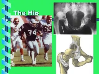
treatment - operative • indications progression of MI, AI decreasing abd
soft tissue lengthening indications for subluxation with reducible hip <8 yrs abd<30 deg MI 25-60% contraindications >4yrs with MI 60-100%
Sp.Quad. Age 18 mo.
2+2yrs Sublux Lt.
Age 2+4 Post op bilat. Adduc,& Prox. Hams. Left only
Age 3+5 13months post op. Lt. Rt. now subluxed Hams. were not released on Rt.
Age 5 +4 2 yrs. Post operative Hams. Release on Rt.
lengthening – post op • spica for 2-3 weeks • abduction orthosis at night
lengthening - results • Cornell • MI <40%, 83% • MI =40%, 23% • MI >60% all failed • AI >27° highly predictive of failure • Age not a significant predictor • Miller • 147 hips • MI <40%, 88% good or fair
bony procedures - recon • >4yrs but before degenerative changes • failed lengthening • <8yrs with MI >40% 12 mo post op
recon options correct deformities, redirect head • femur • varus • derotation • +/- shortening • acetabulum • dega • redirectional
single stage • Soft tissue lengthening • iliopsoas • add longus • add brevis • gracilis • VDRO • peri-ilial acetabuloplasty
single stage - outcomes • 95% stable > 2 yrs • 82% pain free • 14% partial relief • 4% persistent pain
JRP Tracing of x-rays Spastic Diparesis Bilat. Sublux Lt.> Rt.
Pelvis Side View Triradiate Cartilage Osteotomy Location
Osteotome Initial entry Transv. Then curve to Trirad. JRP K-wire
JRP Multiple Osteotomes outline cut & lever laterally, Posterior part 1st
bony procedures - salvage • indications • older pt with painful disloc • options • resection • redirection • interposition • arthrodesis • arthroplasty
complications obturator neurectomy • Hyper-ABD osteonecrosis femoral head • 0 – 11% after reconstruction • femoral osteotomy • increased pressure on the femoral head • during psoas tenotomy
HO • Miller • 192 patients • worse with both hip and spine surgery • prophylaxis • NSAIDS • rads
fracture • bone density • higher risk if prolonged casting
summary • PAIN by late adolescence • few salvage options • GOAL early detection/treatment • ST lengthening <8 yrs, abd<30, MI 25-60 • single stage recon > 4yrs but no degeneration