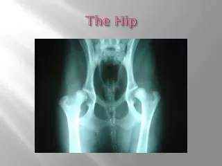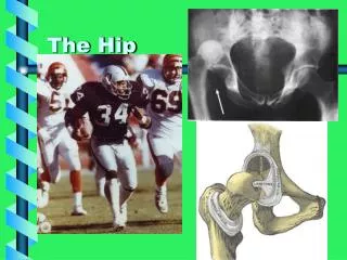
The Hip
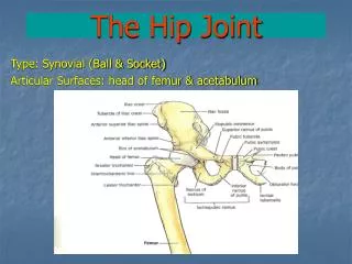
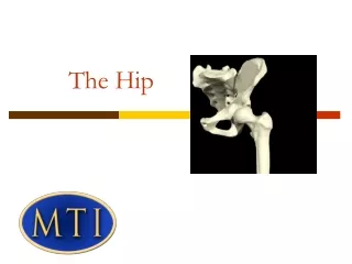
The Hip. Femur Circumduction Acetabulum Iliofemoral External rotation Abduction Pubis Pubofemoral. Flexion Ilium Extension Ischiofemoral Ischium Adduction Internal rotation. The Hip. Anatomy Injuries Evaluation Rehabilitation. Anatomy. Bones Ligaments Muscles.

The Hip
E N D
Presentation Transcript
Femur Circumduction Acetabulum Iliofemoral External rotation Abduction Pubis Pubofemoral Flexion Ilium Extension Ischiofemoral Ischium Adduction Internal rotation
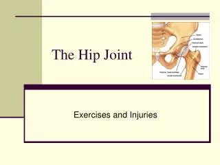
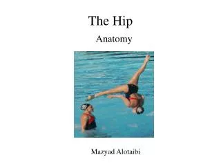
The Hip • Anatomy • Injuries • Evaluation • Rehabilitation
Anatomy • Bones • Ligaments • Muscles
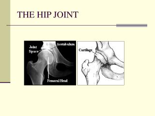
Bones of the hip joint Femur- The ball part of this bone connects to the other three bones to comprise the hip joint. Ilium-This is the largest area of the hip bones. Ischium-The ischium is a curved bone which lies below the ilium. Pubis-The pubis is the front-most area of the hip bones. The ilium, ischium, and pubis come together and form a cup-like shape that the head of the femur fits into. This cup is called the acetabulum.
Ligaments • The iliofemoral ligament attaches the pelvis to the femur. It prevents excessive extension of the hip. • The pubofemoral ligament attaches the pubis bone of the pelvis to the femur. • The posterior of the hip joint capsule is reinforced by the ischiofemoral ligament that attaches from the ischial part of the acetabular rim to the femur.
Motions of the hip • Flexion and extension • Abduction and adduction • Internal and external rotation • Circumduction
Muscles of the hip • Flexor group • Extensor group • External rotators • Internal rotators • Abductors • Adductors
Injuries • Hip flexor strain • Groin strain • Hip pointer • Hip dislocation • MyositisOssificans • ITB syndrome • Stress Fracture • Legg-Calve'-Perthes Disease • Avulsion Fracture • Snapping Hip
Hip pointer • A hip pointer is an injury is to the iliac crest, the bony prominence that can be felt along the waist line. When someone sustains a hip pointer injury, the bone and overlying muscle can be bruised.
Hip dislocation • It requires substantial force to pop the thighbone out of its socket. • In nine out of ten hip dislocations, the head of the thighbone is pushed out and back (posterior dislocation). This leaves the hip in a fixed position, bent and twisted in towards the middle of the body. • http://www.youtube.com/watch?v=VrrpgqwxKoE
Myositis Ossificans • Myositisossificans is an unusual condition that often occurs in athletes who sustain a dirtect blow that causes deep tissue bleeding. • The soft-tissues that were injured in the traumatic event initially develops a hematoma, and subsequently develop the myositisossificans. • The word myositisossificans means that bone forms within the muscle, and this occurs at the site of the hematoma.
ITB syndrome • Iliotibial band syndrome is due to inflammation of the iliotibial band, a thick band of fibrous tissue that runs down the outside of the leg. • The iliotibial band begins at the hip and extends to the outer side of the shin bone (tibia) just below the knee joint. • The band functions in coordination with several of the thigh muscles to provide stability to the outside of the knee joint.
Legg-Calve'-Perthes Disease • LCPD is of unknown origin. It is known that bone death occurs in the ball of the hip due to an interruption in blood flow. • The age of diagnosis is usually between 2 and 12 years old, with the average age of 6. • Most of these children are very active and often very athletic.
Legg-Calve'-Perthes Disease • The first symptoms characterized in LCPD are usually a limp and perhaps pain in the hip, groin, or knee (known as a referred pain). • Often you will first notice limping during your child's active play. • They usually cannot tell you an instance when they hurt themselves.
Avulsion Fracture • Causes: This injury occurs in sports where athletes must start and stop running quickly, must jump, or kick. Such as football, soccer, and basketball. • Signs and Symptoms: You will have sudden pain and may feel a "pop" in your hip or groin. You may have trouble moving your hip and leg. The pain is often worse when the area is touched.
Snapping Hip • The snapping sensation results from the movement of a muscle or tendon over a bony structure. • ITB-When the hip is straight, the band is behind the head of the femur. When the hip bends, the band moves over the head of the femur so that it is then front of it. • Tendons-As you bend the hip, the tendon shifts across the anterior aspect of pelvis; when you straighten the hip, the tendon moves back. • Torn Labrum-If there is a loose flap of cartilage catching within the joint, this may cause a snapping sensation when the hip is moved.