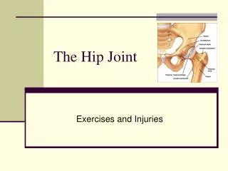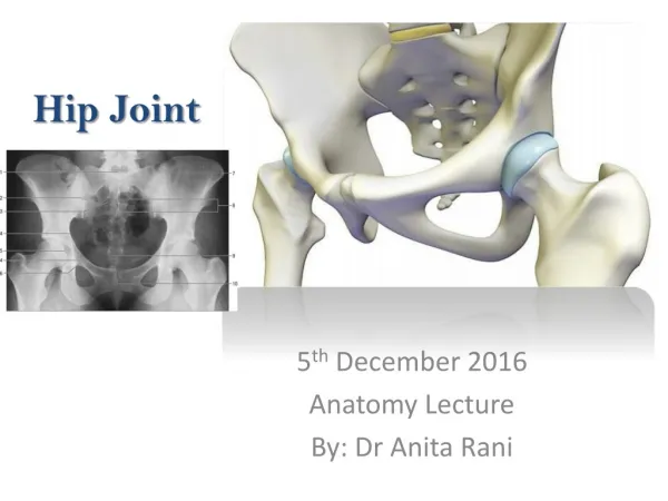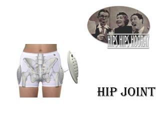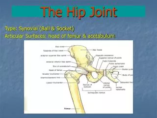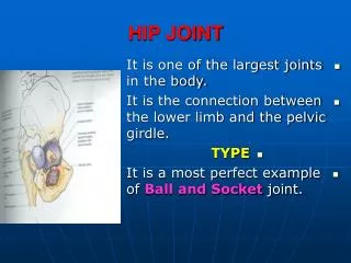
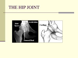
THE HIP JOINT
THE HIP JOINT. Muscles of the Hip. Gluteus Maximus. O: lower posterior iliac crest and posterior surface of the sacrum I: gluteal tuberosity (upper, posterior aspect of the femur) & I.T. band Actions: Extension of the hip External rotation of the hip

THE HIP JOINT
E N D
Presentation Transcript
Gluteus Maximus • O: lower posterior iliac crest and posterior surface of the sacrum • I: gluteal tuberosity (upper, posterior aspect of the femur) & I.T. band • Actions: • Extension of the hip • External rotation of the hip • Lower fibers (below the center of motion) assist in adduction
Gluteus Maximus • Produces hip extension beyond 15 degrees; not used extensively during walking • Strongly used during running, hopping, skipping, and jumping • Best isolated with the knee flexed to reduce hip extension from the hamstrings 40 degrees
Gluteus Medius • O: outer surface of the ilium just below the crest • I: greater trochanter • Actions: • Abduction of the hip • Anterior fibers: Internal rotation, • Posterior fibers: External rotation.
Gluteus Minimus • O: outer surface of the ilium beneath the gluteus medius • I: greater trochanter of the femur • Actions • Abduction of the hip • Internal rotation
Gluteus Medius and Minimus • During walking these muscles abduct (or hold up) the free leg, preventing it from sagging. • Both are important in transferring weight from one leg to the other (e.g. running, hopping, skipping, etc.) • Their effectiveness decreases with age.
Biceps Femoris • Lateral side • Origin: • 1.) Long head - ischial tuberosity; • 2.) Short head - lower half of the linea aspera • Insertion: Head of the fibula • Action: • Extension of hip • External rotation of the hip (and knee) • (Flexion of knee)
Biceps Femoris • One of the hamstring muscles (semitendinosus and semimembranosus) • A two-joint muscle which is a powerful hip extensor unless the knees are flexed • Isolated during leg curls with some external rotation of the hip and knee. This “lines up” the origin and insertion.
Semitendinosus • Medial side; superficial • Origin: Ischial tuberosity • Insertion: Medial surface of proximal end of the tibia • Action: • Extension of the hip • Internal rotation of the hip (and knee) • Flexion of the knee
Semimembranosus • Medial side, deeper than semitendonosus • Origin: Ischial tuberosity • Insertion: Medial surface of the tibia • Action: • Extension of the hip • Internal rotation of the hip (and knee) • Flexion of the knee
Semitendinosus & Semimembranosus • Two-joint muscles • Used in ordinary walking for hip extension • Best exercised with knee flexion exercises (leg curls) with the hip and knee internally rotated • Help to medially stabilize knee
Tensor Fasciae Latae • O: iliac crest • I: iliotibial (I.T.) band • Actions: • Flexion of the hip • Internal rotation • Abduction of the hip
Tensor Fasciae Latae • Prevents external rotation at the hip is flexed Stretching Strengthening
Iliopsoas • Origins: • iliac fossa • vertebral bodies of the last thorasic and lumbar vertebrae • I: lesser trochanter of the femur • Actions: • Flexion of the hip • External rotation
Iliopsoas • Strong hip flexor muscle • Raises legs off the floor from the supine position. • Pulls anteriorly on the lower lumbar vertebrae • May aggravate lower back problems • Abdominal muscles can prevent lumbar strain • Used during complete sit-ups and straight leg sit-ups. • Stretching this muscle requires hyper-extension of the hip.
Pectineous • O: pubic crest or ramus • I: below the linea aspera • Actions • Flexion • Adduction • Internal rotation
Tensor Fasciae Latae Pectineus
Adductor Brevis • Origin: Inferior ramus of pubis • Insertion: Pectineal line (linea aspera) • Actions: • Adduction • External rotation
Adductor Longus • Below the adductor brevis • O: front of the pubis just below its crest • I: middle third of the linea aspera • Actions: • Adduction • Flexion
Adductor Magnus • Located posterior to the longus • O: edge of the pubic crest and ischial tuberosity • I: linea aspera • Actions: • Adduction • External rotation
Gracilis • O: pubic crest • I: medial condyle of tibia • Actions: • Adduction at the hip • Internal rotation • [Flexion at the knee]
Adductor Muscles • Adductor Brevis • Adductor Longus • Adductor Magnus • Gracilis • Not heavily used in ordinary movements • Horse back riding, the breaststroke kick in swimming
Sartorius • Origin: Anterior-superior spine of the ilium • Insertion: Anterior medial condyle of the tibia (behind the medial condyle) • Action: • Flexion of hip • External rotation of the hip • [Flexion of the knee]
Sartorius • Longest muscle in the body • It is a two-joint muscle; hip flexion and knee flexion • It is weak when both actions take place at the same time.
Rectus Femoris • Two joint muscle; most superficial • Origin: anterior-inferior iliac spine of the ilium • Insertion: top of the patella and patellar ligament to the tibial tuberosity • Actions: • Flexion of the hip [Extension of the knee]
Rectus Femoris • A two-joint muscle: hip flexion and knee extension • Powerful knee extension when the hip is extended but weaker when the hip is flexed. • The gluteus maximus and the hamstring muscles will extend the hip making the rectus femoris stronger during knee extension
Hip Rotator Muscles • ANTERIOR • Obturator Externus Anterior
Hip Rotator Muscles • POSTERIOR • Piriformis • Gemellus superior • Obturator internus Gemellus inferior • Quadratus femoris Posterior
Hip Rotator Muscles Posterior Posterior
Hip Rotator Muscles Posterior Anterior
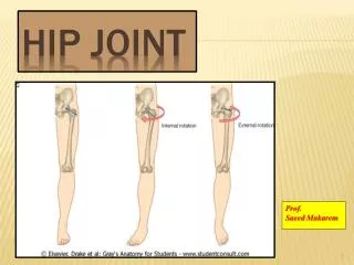
Six Hip Rotator Muscles • Common action is External Rotation • Powerful external rotation of the hip is required to throw a baseball, swing a bat or golf club. • The sciatic nerve passes just inferior to the piriformis therefore a tight piriformis muscle my contribute to compression on the sciatic nerve.
Name the action at her hip Abduction
Name the action at his right hip Flexion
Name the actions at her hip Extension, Abduction & External Rotation
Name the two action at his right hip Extension and External Rotation
Name the action at his hip Flexion
Name the actions at his hip Flexion and External Rotation and Abduction
Name the actions at his hip Flexion and Adduction
3. The Femur 1. 2. 1 = 2 = 3 = 4 = 5 = 6 = 7 = Head Neck Greater trochanter Lesser trochanter Linea Aspera Medial condyle Lateral condyle 4. 5. 6. 7.