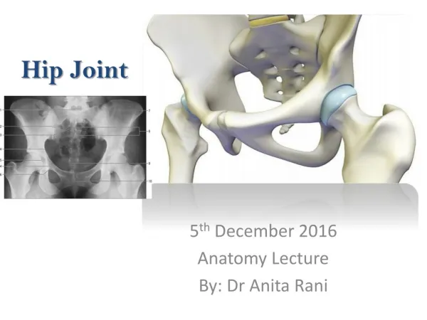
Hip Joint
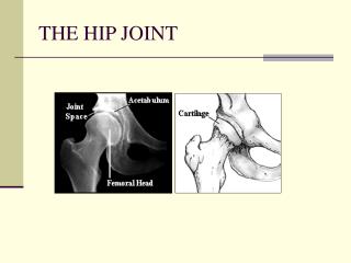
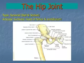
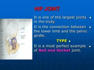
Hip Joint. Ball and socket joint of synovial type . Connects the pelvic girdle to the lower limb Made up of femoral head and acetabulum Designed for stability and wide range of movement Covered with a thin layer of hyaline cartilage.

Hip Joint
E N D
Presentation Transcript
Ball and socket joint of synovial type. • Connects the pelvic girdle to the lower limb • Made up of femoral head and acetabulum • Designed for stability and wide range of movement • Covered with a thin layer of hyaline cartilage
The articular surface is horse-shoe shaped and is deficient inferiorly at the acetabular notch • Acetabular notch is covered by acetabular ligament • Has a labrum whichis a circular layer of cartilage which surrounds the outer part of the acetabulum making the socket deeper and so helping provide more stability • Acetabularlabral tears are a common injury from major or repeated minor trauma
Capsule of the Hip Joint • The capsule is tough connective tissue • It is attached to the margins of the labrum • It forms a sleeve extends between the margins of acetabulum and the neck of femur • It supports the joint
The synovial Membrane • It is a thin membrane • It lines the capsule and covers intracapsular areas a way from the articular surfaces • It secrets the synovial fluid for lubrication and articular cartilage nourishment
Ligaments of the Hip Joint Iliofemoral ligament: Connects the pelvis to the femur anteriorly It resembles a Y in shape From inferior iliac spine and rim of acetabulum To intertrochantaric line Stabilises the hip by limiting hyperextension 10o-20o Pubofemoral Ligament: From obturator crest of pubic bone Prevents over abduction Ischiofemoral Ligament: From ischial part of acetabulum To the neck of femur poateriorly
Ligaments of the Hip Joint Cont., • Transverse acetabular Ligament: Bridges acetabular notch. • Ligament of head of femur: Flat and triangular in shape Lies within joint, ensheathed by synovial membrane
Nerve Supply of the Hip Joint • Femoral (L2,3,4) • Obturator(L2, 3, 4) • Sciatic (L4,5, S1, 2,) • Nerve to quadratusfemoris • Superior gluteal nerve • Referred pain to the knee can hide hip pathology and visa versa
Blood Supply of the Hip Joint • Femoral Artery Supplies the capsule and neck of femur It gives: Medial circumflex femoral artery Lateral circumflex femoral artery • Obturator artery Supplies the head of femur • Avascular necrosis
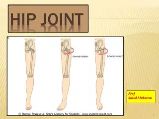
Movements of the Hip Joint Multiaxial • Flexion/extension • Abduction/adduction • Internal/external rotation
Muscles Acting on the Hip • Flexors: Iliopsoas, sartorius, tensor fascia lata, rectus femoris,pectineus, adductor longus, brevis, and magnus, gracilis • Extensors: hamstrings, addcutormagnus, gluteus maximus • Adductors: adductor longus, brevis, and magnus, gracilis, pectineus, obturatorinternus • Abductors: gluteus medius, minimus, tensor fascia lata • External rotators: obturatorexternus, internus, piriformis, quadratusfemoris, gluteus maximus • Internal Rotators: gluteus medius, gluteus minimus, tensor fascia lata.
a Flexion/Extension Abduction/Adduction Internal/External rotation
Stability of the Hip Joint It is the most stable joint due to: • Size, strength and position of joint ligaments • Medial and lateral rotators • Muscles and ligaments pull the head towards the acetabulum • The acetabulum is deepened by the labrum Dislocation is rare • Posterior dislocation • Congenital hip dislocation • Hip replacement
Anterior Group They include: • Quadriceps muscle Rectus femoris Vastusmedialis Vastusintermedius Vastuslateralis • All arise from front and sides of femur and rectus femoris arises from above the acetabulum • They are inserted in a common tendon in tibial tuberosity • Nerve supply is the femoral nerve • Action: they extend the knee joint in addition the rectus head flexes the hip
Anterior Group Cont., Sartorius • Origin: anterior superior iliac spine • Insertion: medial side of tibia • Nerve supply: femoral nerve • Action: flexion of hip and knee joints • Iliotibialtract: • Origin:iliac crest • Insertion: lateral site of tibia • Nerve supply: femoral nerve • Action: abduction of the hip
Anterior Group Cont., Iliopsoas: • Consists of 2 parts Iliacus • Origin: iliac fossa Psoas major • Origin: transverse processes of lumbar vertebrae • Both muscles inserted to lesser trochanter • Action: they are primary hip flexor • Nerve supply: Lumbar spinal nerves and femoral nerve
Medial Group • They are called adductor group • They include: Adductor longus, brevis, and magnus Gracilis Pectenius • All muscles arise from pubic and ischial borders • Pectenius arises from superior pubic ramus • All are inserted to the back of femur while the magnus extends into adductor tubercle on the medial condyle • Nerve supply is obturator nerve • Action is adduction and medial rotation
Posterior Group • They are called hamsrings muscles • They include: Semimembranosus Semitendinosus Biceps femoris Adductor magnus • They arise from the ischial tuberosity • Semimembranosus and semitendinosus inserted on the medial side of tibia • Biceps femoris inserted into the head of fibula • Adductor magnus inserted into the back of femur • Nerve supply is sciatic nerve • Action is extension of the hip and flexion of knee joint
4. Hamstrings • 5. semitendinosus • 6. semimembranosus • 7. Biceps femoris • 8. Adductor magnus