HIP Joint
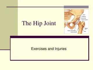
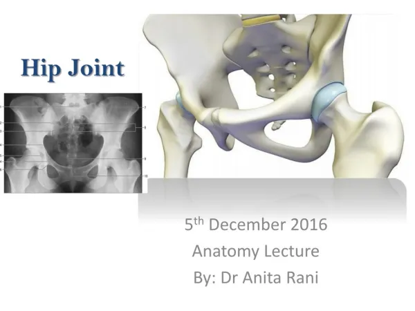
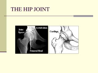
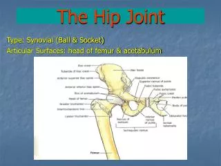
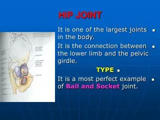
HIP Joint. The hip joint is the articulation between the femur and the acetabulum of the pelvis and its primary function is to support the weight of the body in both static (e.g. standing) and dynamic (e.g. walking or running) postures .

HIP Joint
E N D
Presentation Transcript
The hip joint is the articulation between the femur and the acetabulum of the pelvis and its primary function is to support the weight of the body in both static (e.g. standing) and dynamic (e.g. walking or running) postures
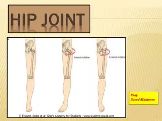
Seven different kinds of movements are possible in the hip joint: • Flexion and extension • Abduction and adduction • Internal (medial) and external (lateral) rotation • Circumduction • A synovial joint that can produce movement in more than one axis is called a multiaxial joint
The large head of the femur is completely covered in hyaline cartilage except for a small area called the fovea or pit. This is the site of attachment for an intracapsular ligament [ligament of head of femur] that attaches directly from the head of the femur to the acetabulum. • There is also a small ligament called ligamentum teres or the ligament of the head of the femur. This structure is not that important as a ligament but can often be vitally important as a conduit of a small artery to the head of the femur. This arterial branch is not present in everyone but can become the only blood supply to the bone in the head of the femur when the neck of the femur is fractured or disrupted by injury in childhood.
Ligaments • At the front of the joint, the strong iliofemoral ligament attaches from the pelvis to femur. This Y-shaped ligament is also known as the ligament of Bigelow. This ligament seeks to resist excessive extension of the hip joint. It is often considered to be the strongest ligament in the human body. • The pubofemoral ligament attaches across the front of the joint from the pubis bone of the pelvis to the femur. This ligament is orientated more inferiorly than the iliofemoral ligament and reinforces the inferior part of the hip joint capsule. It also blends with the medial parts of the iliofemoral ligamemt. • The posterior of the hip joint capsule is reinforced by the ischiofemoral ligament that attaches from the ischial part of the acetabular rim to the femur.
LabrumThough not actually a ligament, the acetabular labrum (latin for “lip”) contributes to the stability of the hip joint by encompassing more than half of the femoral head. Together with the transverse acetabular ligament, it creates a deep spherical "cup".
Muscles producing movements at the hip joint • The 17 muscles that cause movement in the hip can be divided into four groups according to their orientation around the hip joint: the gluteal group, the lateral rotator group, the adductor group, and the iliopsoas group. These muscles produce flexion, extension, lateral rotation, medial rotation, abduction and adduction. • Many of the hip muscles are responsible for more than one type of movement in the hip, as different areas of the muscle act on tendons in different ways.
Gluteal group • The gluteal muscles include the gluteus: maximus, medius & minimus, and tensor fasciae latae. They cover the lateral surface of the ilium. • The gluteus maximus, which forms most of the muscle of the buttock, originates primarily on the ilium and sacrum and inserts on the gluteal tuberosity of the femur as well as the iliotibial tract, a strong fibrous tissue that runs along the lateral thigh to the tibia & fibula. • The gluteus medius and gluteus minimus originate anterior to the gluteus maximus on the ilium and both insert on the greater trochanter of the femur. • The tensor fasciae latae shares its insertion with the gluteus maximus at the ilium and also shares the insertion at the iliotibial tract.
Adductor group • The adductor brevis, adductor longus, adductor magnus, pectineus, and gracilis make up the adductor group. The adductors all orginate on the pubis and insert on the medial, posterior surface of the femur, with the exception of the gracilis which inserts just below the medial condyle of the tibia.
Iliopsoas group • The iliacus and psoas major comprise the iliopsoas group. The iliopsoas is a large muscle that runs from the transverse processes of the T-12 to L-5 vertebrae, joins with the iliacus via its tendon, and connects to the lesser trochanter of the femur. The iliacus originates on the iliac fossa of the ilium. Together these muscles are commonly referred to as the "iliopsoas".
Lateral rotator group • This group consists of the externus and internus obturators, the piriformis, the superior and inferior gemelli, and the quadratus femoris. These six originate at or below the acetabulum of the ilium and insert on or near the greater trochanter of the femur.