Download
1 / 67
700 likes | 1.51k Vues
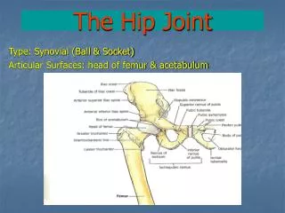
The Hip Joint and Pelvic Girdle. Anatomy and Kinesiology 420:024. Objectives. Bones, bony landmarks and joints Muscles Movements. Hip Joint. Hip joint (femur and acetabulum of pelvis) Relatively stable due to: Bony architecture Strong ligaments Large supportive muscles

E N D
The Hip Joint and Pelvic Girdle Anatomy and Kinesiology 420:024
Objectives • Bones, bony landmarks and joints • Muscles • Movements
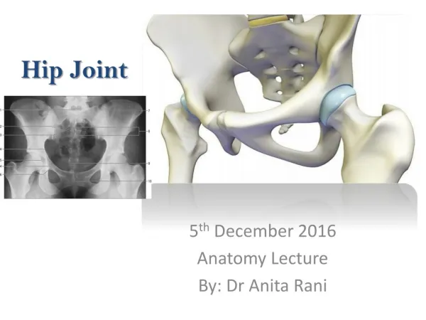
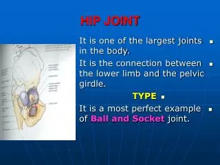
Hip Joint • Hip joint (femur and acetabulum of pelvis) • Relatively stable due to: • Bony architecture • Strong ligaments • Large supportive muscles • Hip joint and pelvic girdle work together much like shoulder joint and girdle • Pelvis moves in response to trunk and/or thigh movement • Other bones to consider • Tibia • Fibula • Patella
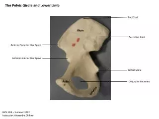
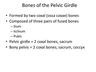
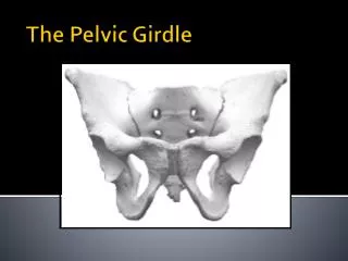
Pelvic Girdle • Pelvic girdle Pelvic bones, sacrum, coccyx • Pelvic bones: • Each pelvic bone is made up of three fused bones ilium, ischium, & pubis • Right & left pelvic bone joined together posteriorly by sacrum • Sacrum: • Extends from spinal column with 5 fused vertebrae • Coccyx: • Extends posteriorly from sacrum with 3 fused vertebrae
Lateral femoral epicondyle Medial femoral epicondyle Patella Lateral tibial condyle Medial tibial condyle Head of fibula Tibial tuberosity
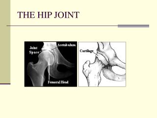
Joints • Hip joint • AKA Acetabular femoral joint • Diarthrodial multiaxial ball and socket • Movements • Planes and axes
Joints • Pubic symphisis • Amphiarthrodial cartilagenous joint • Slightly moveable
Joints • Sacroiliac joints • Diarthrodial gliding joints • Slightly moveable
Objectives • Bones, bony landmarks and joints • Muscles • Movements
Anterior: Iliopsoas Sartorius Rectus femoris Tensor fasciae latae Posterior: Gluteus maximus Biceps femoris Semitendonosus Semimembranosus Deep 6 external rotators Muscles • Lateral: • Gluteus medius • Gluteus minimus • Medial: • Adductor brevis • Adductor longus • Adductor magnus • Gracilis • Pectineus
Anterior: Iliopsoas Sartorius Rectus femoris Tensor fasciae latae Posterior: Gluteus maximus Biceps femoris Semitendonosus Semimembranosus Deep 6 external rotators Muscles • Lateral: • Gluteus medius • Gluteus minimus • Medial: • Adductor brevis • Adductor longus • Adductor magnus • Gracilis • Pectineus
Anterior: Iliopsoas Sartorius Rectus femoris Tensor fasciae latae Posterior: Gluteus maximus Biceps femoris Semitendonosus Semimembranosus Deep 6 external rotators Muscles • Lateral: • Gluteus medius • Gluteus minimus • Medial: • Adductor brevis • Adductor longus • Adductor magnus • Gracilis • Pectineus
Anterior: Iliopsoas Sartorius Rectus femoris Tensor fasciae latae Posterior: Gluteus maximus Biceps femoris Semitendonosus Semimembranosus Deep 6 external rotators Muscles • Lateral: • Gluteus medius • Gluteus minimus • Medial: • Adductor brevis • Adductor longus • Adductor magnus • Gracilis • Pectineus
Objectives • Bones, bony landmarks and joints • Muscles • Movements
Movements • Flexion • Movement of femur straight anteriorly • Extension • Movement of femur straight posteriorly
Movements • Abduction • Movement of femur laterally to side away from midline • Adduction • Movement of femur medially toward midline
Movements • Horizontal adduction • Movement of femur in a horizontal or transverse plane toward the midline • Horizontal abduction • Movement of femur in a horizontal or transverse plane away from the midline
Movements • External rotation • Movement of femur laterally around its long axis away from midline • Internal rotation • Movement of femur medially around its long axis toward midline
Movements • Diagonal abduction • Movement of femur in a diagonal plane away from midline of body • Diagonal adduction • Movement of femur in a diagonal plane toward midline of body
Movements • Anterior pelvic tilt • Anterior movement of upper pelvis; iliac crest tilts forward in a sagittal plane • Posterior pelvic tilt • Posterior movement of upper pelvis; iliac crest tilts backward in a sagittal plane
Movements • Left lateral pelvic tilt • Left pelvis moves inferiorly in relation to right pelvis in frontal plane • Right lateral pelvic tilt • Right pelvis moves inferiorly in relation to left pelvis in frontal plane
Movements • Left transverse pelvic tilt • Left pelvis moves posteriorly in relation to the right in transverse plane • Right transverse pelvic tilt • Right pelvis moves posteriorly in relation to the left in transverse plane
FLEXION • Superior movement of the femur in the sagittal plane
FLEXION • Iliopsoas • Rectus femoris • Sartorius • Tensor fasciae latae • Pectineus
EXTENSION • Inferior movement of the femur in the sagittal plane