Download
1 / 53
570 likes | 964 Vues
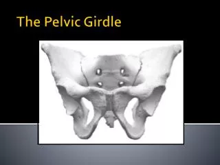
Bones of the Pelvic Girdle. Formed by two coxal (ossa coxae) bones Composed of three pairs of fused bones Ilium Ischium Pubis Pelvic girdle = 2 coxal bones, sacrum Bony pelvis = 2 coxal bones, sacrum, coccyx. Bones of the Pelvic Girdle.

E N D
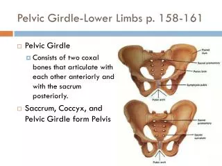
Bones of the Pelvic Girdle Formed by two coxal (ossa coxae) bones Composed of three pairs of fused bones Ilium Ischium Pubis Pelvic girdle = 2 coxal bones, sacrum Bony pelvis = 2 coxal bones, sacrum, coccyx
Bones of the Pelvic Girdle The total weight of the upper body rests on the pelvis It protects several organs Reproductive organs Urinary bladder Part of the large intestine
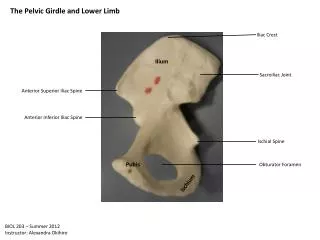
lliac crest Sacroiliac joint llium Coxal bone (or hip bone) Sacrum Pelvic brim Coccyx Ischial spine Pubis Acetabulum Pubic symphysis Ischium Pubic arch (a) Figure 5.26a
IIium Ala IIiac crest Posterior superior iliac spine Anterior superior iliac spine Posterior inferior iliac spine Anterior inferior iliac spine Greater sciatic notch Acetabulum Ischial body Body of pubis Ischial spine Pubis Ischial tuberosity Inferior pubic ramus Ischium Obturator foramen Ischial ramus (b) Figure 5.26b
Gender Differences of the Pelvis The female inlet is larger and more circular The female pelvis as a whole is shallower, and the bones are lighter and thinner The female ilia flare more laterally The female sacrum is shorter and less curved The female ischial spines are shorter and farther apart; thus the outlet is larger The female pubic arch is more rounded because the angle of the pubic arch is greater
False pelvis Inlet of true pelvis Pelvic brim Pubic arch (less than 90°) False pelvis Inlet of true pelvis Pelvic brim Pubic arch (more than 90°) (c) Figure 5.26c
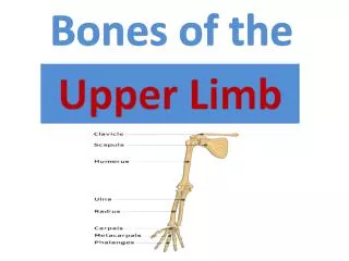
Bones of the Lower Limbs Femur—thigh bone The heaviest, strongest bone in the body Proximal end articulation Head articulates with the acetabulum of the coxal (hip) bone Distal end articulation Lateral and medial condyles articulate with the tibia in the lower leg
Neck Head Inter- trochanteric line Lesser trochanter Lateral condyle Patellar surface (a) Figure 5.27a
Greater trochanter Head Inter- trochanteric crest Lesser trochanter Gluteal tuberosity Intercondylar fossa Medial condyle Lateral condyle (b) Figure 5.27b
Bones of the Lower Limbs The lower leg has two bones Tibia—Shinbone; larger and medially oriented Proximal end articulation Medial and lateral condyles articulate with the femur to form the knee joint Fibula—Thin and sticklike; lateral to the tibia Has no role in forming the knee joint
Intercondylar eminence Medial condyle Lateral condyle Head Tibial tuberosity Proximal tibiofibular joint Interosseous membrane Anterior border Fibula Tibia Distal tibiofibular joint Medial malleolus Lateral malleolus (c) Figure 5.27c
Bones of the Lower Limbs The foot Tarsals—seven bones Two largest tarsals Calcaneus (heel bone) Talus Metatarsals—five bones form the sole of the foot Phalanges—fourteen bones form the toes
Phalanges: Distal Middle Proximal Tarsals: Medial cuneiform Metatarsals Tarsals: Intermediatecuneiform Lateral cuneiform Navicular Cuboid Talus Calcaneus Figure 5.28
Arches of the Foot Bones of the foot are arranged to form three strong arches Two longitudinal One transverse
Medial longitudinal arch Transverse arch Lateral longitudinal arch Figure 5.29
Joints Articulations of bones Functions of joints Hold bones together Allow for mobility Two ways joints are classified Functionally Structurally
Functional Classification of Joints Synarthroses Immovable joints Amphiarthroses Slightly moveable joints Diarthroses Freely moveable joints
Structural Classification of Joints Fibrous joints Generally immovable Cartilaginous joints Immovable or slightly moveable Synovial joints Freely moveable
Fibrous Joints Bones united by collagenic fibers Types Sutures Immobile Syndesmoses Allows more movement than sutures but still immobile Example: Distal end of tibia and fibula Gomphosis Immobile
Fibrous joints Fibrous connective tissue (a) Suture Figure 5.30a
Fibrous joints Tibia Fibula Fibrous connective tissue (b) Syndesmosis Figure 5.30b
Cartilaginous Joints Bones connected by cartilage Types Synchrondrosis Immobile Symphysis Slightly movable Example: Pubic symphysis, intervertebral joints
Cartilaginous joints First rib Hyaline cartilage Sternum (c) Synchondrosis Figure 5.30c
Cartilaginous joints Vertebrae Fibrocartilage (d) Symphysis Figure 5.30d
Cartilaginous joints Pubis Fibro- cartilage (e) Symphysis Figure 5.30e
Synovial Joints Articulating bones are separated by a joint cavity Synovial fluid is found in the joint cavity
Synovial joints Scapula Articular capsule Articular (hyaline) cartilage Humerus (f) Multiaxial joint (shoulder joint) Figure 5.30f
Synovial joints Humerus Articular (hyaline) cartilage Articular capsule Radius Ulna (g) Uniaxial joint (elbow joint) Figure 5.30g
Synovial joints Ulna Radius Articular capsule Carpals (h) Biaxial joint (intercarpal joints of hand) Figure 5.30h
Features of Synovial Joints Articular cartilage (hyaline cartilage) covers the ends of bones Articular capsule encloses joint surfaces and lined with synovial membrane Joint cavity is filled with synovial fluid Reinforcing ligaments
Structures Associated with the Synovial Joint Bursae—flattened fibrous sacs Lined with synovial membranes Filled with synovial fluid Not actually part of the joint Tendon sheath Elongated bursa that wraps around a tendon
Acromion of scapula Ligament Joint cavity containing synovial fluid Bursa Ligament Articular (hyaline) cartilage Tendon sheath Synovial membrane Fibrous layer of the articular capsule Tendon of biceps muscle Humerus Figure 5.31
Nonaxial Uniaxial Biaxial Multiaxial (a) Plane joint (a) Figure 5.32a
Nonaxial Uniaxial Biaxial Multiaxial (b) Humerus Ulna (b) Hinge joint Figure 5.32b
Nonaxial Uniaxial Biaxial Multiaxial Ulna (c) Radius (c) Pivot joint Figure 5.32c
Nonaxial Uniaxial Biaxial Multiaxial (d) Metacarpal Phalanx (d) Condylar joint Figure 5.32d
Nonaxial Uniaxial Biaxial Multiaxial Carpal Metacarpal #1 (e) (e) Saddle joint Figure 5.32e
Nonaxial Uniaxial Biaxial Multiaxial (f) Head of humerus Scapula (f) Ball-and-socket joint Figure 5.32f
Inflammatory Conditions Associated with Joints Bursitis—inflammation of a bursa usually caused by a blow or friction Tendonitis—inflammation of tendon sheaths Arthritis—inflammatory or degenerative diseases of joints Over 100 different types The most widespread crippling disease in the United States Initial symptoms: pain, stiffness, swelling of the joint
Clinical Forms of Arthritis Osteoarthritis Most common chronic arthritis Probably related to normal aging processes Rheumatoid arthritis An autoimmune disease—the immune system attacks the joints Symptoms begin with bilateral inflammation of certain joints Often leads to deformities
Clinical Forms of Arthritis Gouty arthritis Inflammation of joints is caused by a deposition of uric acid crystals from the blood Can usually be controlled with diet More common in men
Developmental Aspects of the Skeletal System At birth, the skull bones are incomplete Bones are joined by fibrous membranes called fontanels Fontanels are completely replaced with bone within two years after birth
Parietal bone Frontal bone of skull Occipital bone Mandible Clavicle Scapula Radius Ulna Humerus Femur Tibia Ribs Vertebra Hip bone Figure 5.34
Skeletal Changes Throughout Life Fetus Long bones are formed of hyaline cartilage Flat bones begin as fibrous membranes Flat and long bone models are converted to bone Birth Fontanels remain until around age 2
Skeletal Changes Throughout Life Adolescence Epiphyseal plates become ossified and long bone growth ends Size of cranium in relationship to body 2 years old—skull is larger in proportion to the body compared to that of an adult 8 or 9 years old—skull is near adult size and proportion Between ages 6 and 11, the face grows out from the skull
Skeletal Changes Throughout Life Curvatures of the spine Primary curvatures are present at birth and are convex posteriorly Secondary curvatures are associated with a child’s later development and are convex anteriorly Abnormal spinal curvatures (scoliosis and lordosis) are often congenital