Download
1 / 31
320 likes | 618 Vues
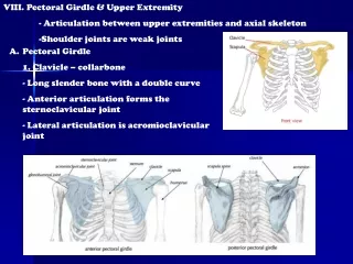
BONES OF THE PECTORAL GIRDLE AND UPPER EXTREMITY . Right clavicle superior view. Conoid tubercle. Shaft. (Posterior end). (Anterior end). Sternal (medial) end. Acromial (lateral) end. Left scapula posterior view. Superior angle. Supraspinous fossa. Medial border. Acromion.

E N D
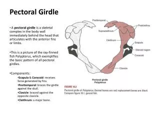
BONES OF THE PECTORAL GIRDLE AND UPPER EXTREMITY
Right clavicle superior view Conoid tubercle Shaft (Posterior end) (Anterior end) Sternal (medial) end Acromial (lateral) end
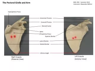
Left scapula posterior view Superior angle Supraspinous fossa Medial border Acromion Spine Infraspinous fossa Coracoid process Lateral border Inferior angle Lateral angle Glenoid cavity
Left scapula anterior view Superior angle Superior border Suprascapular notch Medial border Acromion process Subscapular fossa Lateral border Coracoid process Inferior angle Lateral angle
Right humerus anterior view Head of humerus Greater tubercle Anatomic neck Intertubercular sulcus Place* where deltoid muscle “attaches” (deltoid tuberosity) *Radial groove runs obliquely on posterior side of shaft Surgical neck Lesser tubercle Coronoid fossa Radial fossa Medial epicondyle Capitulum Trochlea
Proximal right humerus – anterior view Intertubercular sulcus Greater tubercle Surgical neck Lesser tubercle Anatomical neck Head of humerus
Distal right humerus – anterior view Lateral epicondyle Radial fossa (site where headofradius “fits” when elbow is flexed) Capitulum (attaches to headof radius) Trochlea (attaches to trochlear notchofulna) Coronoid fossa (site where coronoidprocess ofulna “fits” when elbow is flexed) Medial epicondyle
Distal right humerus – posterior view Medial epicondyle Trochlea Capitulum Lateral epicondyle Olecranon fossa (site where olecranonprocess ofulna “fits” when elbow is extended)
Left ulna lateral view Trochlear notch Coronoid process Radial notch Head of ulna Olecranon process
Proximal left ulna lateral view Coronoid process (“fits” into coronoid fossaof distal humerus) Trochlear notch Olecranon process (“fits” into olecranon fossaof distal humerus) Radial notch (“fits” into headofradius)
Distal left ulna medial view Head of ulna Styloid process of ulna
Left radius anterior view Distal, middle, and proximal phalanges* *Thumb = no middle Head of radius Neck of radius Styloid process of radius 4 Radial tuberosity (Proximal end) (Distal end) D Metacarpals 8 carpal bones
Right hand anterior view Left radius anterior view Distal, middle, and proximal phalanges* *Thumb = no middle 1 Head of radius Neck of radius Styloid process of radius 2 3 4 Radial tuberosity 5 (Proximal end) (Distal end) D Metacarpals 8 carpal bones
8 Carpal bones – Right hand – anterior view Trapezoid Pisiform Hamate Capitate Trapezium Triquetral Lunate Scaphoid “Sally Left The Party To Take Cathy Home”
Male pelvis – ilium, ischium, pelvis – anterior view Iliac crest Sacroiliac joint Anterior superior iliac spine Iliac fossa Arcuate line (pelvic brim) Anterior inferior iliac spine Superior ramus of pubis Acetabulum Obturator foramen Pubic arch
Male pelvis – ilium, ischium, pelvis – posterior view Posterior superior iliac spine Posterior inferior iliac pine Greater sciatic notch ischial spine Lesser sciatic notch ischial tuberosity ischial ramus
Male pelvis – ilium, ischium, pelvis – anterior view Pubic crest Pubic symphysis Superior ramus of pubis Acetabulum Inferior ramus of pubis ischial tuberosity
Right femur Lateral condyle Patellar surface Medial epicondyle Medial condyle
Right femur Gluteal tuberosity Medial condyle* Linea aspera Intercondylar fossa Lateral condyle* Lateral epicondyle *Condyles connect with condylesoftibia
Right proximal anterior femur Greater trochanter Head of femur Intertrochanteric line Neck Lesser trochanter Fovea capitis
Right proximal posterior femur Head of femur Lesser trochanter Neck Intertrochanteric crest Greater trochanter
The following are important • special structures of the tibia: • Lateral condyle • Medial condyle • Intercondylar eminence • Tibial tuberosity • Anterior crest • Medial malleolus
Lateral condyle Medial condyle Tibial tuberosity Anterior crest Medial malleolus
Head Interosseus border (crest) Lateral malleolus
Right foot posterior view Medial Malleolus (tibia) Lateral malleolus* (fibula) Talus *Head of the fibula is at the proximal end Calcaneus
Right foot lateral view Tibia Talus (tarsal bone) Navicular (tarsal bone) Lateral malleolus (fibula) Intermediate cuneiform (tarsal bone) Calcaneus (tarsal bone) 1 2 3 Cuboid (tarsal bone) 4 5 Lateral cuneiform (tarsal bone) Metatarsals Phalanges
Left foot superior/lateral view Calcaneus Navicular (tarsal bone) Talus Medial Intermediate Lateral Cuboid (tarsal bone) Cuneiform 1 2 3 4 5 Metatarsals
Left foot *Great toe = no middle phalanx Distal, middle, and proximal phalanges*