Download
1 / 43
440 likes | 559 Vues
Learn about the pelvic girdle (hip bone) and lower limbs organization, bones landmarks, hip structure, and the anatomy of the femur in human skeletal system.

E N D
Appendicular SkeletonPelvic Girdle & Lower limbs Dr. NabilKhouri MD, MSc, Ph.D
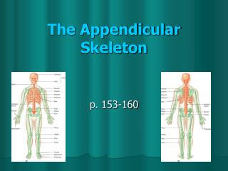
Organization of the Lower Limb • It is divided into: • The Gluteal region • The thigh • The knee • The leg • The ankle • The foot • The thigh and the leg have compartments with its own muscles that perform group functions and its own distinct nerve & blood supply
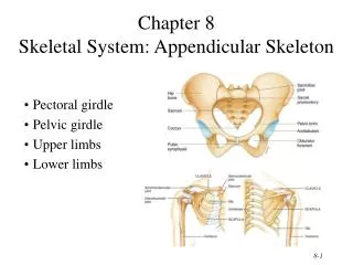
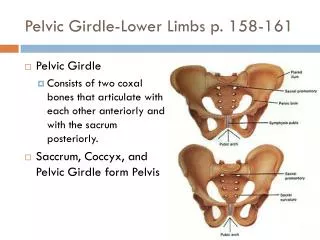
Hip Bone: • The mature hip bone is the large, flat pelvic boneformed by the fusion of three primary bones. • Ilium, • Ischium, and • Pubis • The three separate bones are joined by cartilage at the acetabulum. • At puberty, these three bones fuse together to form one large, irregular bone. • The hip bones articulate with the sacrum at the sacroiliac joints and form the anterolateral walls of the pelvis; • They also articulate with one another anteriorly at the symphysis pubis.
Bones Landmarks of the Gluteal Region • Ilium • Iliac crest • Anterior superior & inferior iliac spines • Posterior superior & inferior iliac spines • Greater sciatic notch • Ischium • Body • Ramus • Ischial spine • Ischial tuberosity • Greater & lesser sciatic notches/foramina • Pubis • Body • Superior & inferior rami • Symphysis pubis • Obturator foramen/membrane • Pubic crest/tubercle
Ilium: • Anteriorly, The ilium is separated into upper and lower parts by a rounded ridge on the medial surface termed THE ARCUATE LINE. • Posteriorly, the ridge is sharp and lies immediately superior to the surface of the bone that articulates with the sacrum. • THE BODY OF THE ILIUM joins the pubis and ischium to form the acetabulum. • Anteriorly, the ilium has stout ANTERIOR SUPERIOR AND ANTERIOR INFERIOR ILIAC SPINES that provide attachment for ligaments and tendons of lower limb muscles. • Beginning at the anterior superior iliac spine (ASIS), the long curved and thickened superior border of the ala of the ilium, THE ILIAC CREST, extends posteriorly, terminating at the posterior superior iliac spine. • A prominence on the external lip of the crest, THE TUBERCLE OFTHE ILIAC CREST (ILIAC TUBERCLE), lies 5–6 cm posterior to the ASIS. • The posterior inferior iliac spine marks the superior end of the greater sciatic notch. • THE LATERAL POSTERIOR SURFACE OF THE ALA OF THE ILIUM has three rough curved lines—the POSTERIOR, ANTERIOR, AND INFERIOR GLUTEAL LINES—that demarcate the proximal attachments of the three large gluteal muscles. • Medially, each ala has a large, smooth depression, the iliac fossa
Ischium • The superior part of the BODY OF THE ISCHIUM fuses with the pubis and ilium, forming the postero-inferior aspect of the acetabulum. • The RAMUS OF THE ISCHIUM joins the inferior ramusof the PUBISto form a bar of bone, the ISCHIOPUBIC RAMUS. • The posterior border of the ischium forms the inferior margin of a deep indentation called the GREATER SCIATIC NOTCH. • The large, triangular ISCHIAL SPINE at the inferior margin of this notch provides ligamentous attachment. • The rough bony projection at the junction of the inferior end of the body of the ischium and its ramus is the large ISCHIAL TUBEROSITY.
Pubis • The pubis is divided into a flattened medially placed BODYand SUPERIOR AND INFERIOR RAMI that project laterally from the body. • Medially, the SYMPHYSIAL SURFACE of the body of the pubis articulates with the corresponding surface of the body of the contralateral pubis by means of the pubic symphysis. • The anterosuperior border of the united bodies and symphysis forms the PUBIC CREST. • Small projections at the lateral ends of this crest, the PUBICTUBERCLES. • The posterior margin of the superior ramus of the pubis has a sharp raised edge, the PECTEN PUBIS.
Foramina: • Greater Sciatic Foramen • Lesser Sciatic Foramen • Obturator Foramen
The Acetabulum The acetabulum is formed by the pubis, ischium and ilium bones
Male skeleton larger and heavier Heart shape Angle formed between 2 arms of pubic 50-60 The ischial spine project medially larger articular surfaces larger muscle attachments Female pelvis wider & shallower Circular Angle between 2 arms of pubic 80-85 The ischial spine project less medially larger pelvic inlet & outlet Female and Male Skeletons
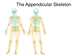
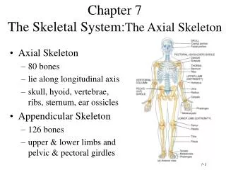
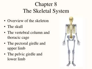
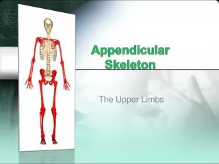
Lower Limbs • The three segments of the lower limb are the thigh, leg, and foot • They carry the weight of the erect body, and are subjected to exceptional forces when one jumps or runs
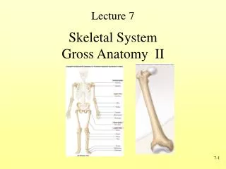
Femur • The sole bone of the thigh is the femur, the largest and strongest bone in the body • It articulates proximally with the hip and distally with the tibia and fibula • Major markings include the head, fovea capitis, greater and lesser trochanters, gluteal tuberosity, lateral and medial condyles and epicondyles, linea aspera, patellar surface, and the intercondylar notch
Superior (Proximal) End • Its proximal end is characterized by • Head • Neck, • Two large projections (the greater and lesser trochanters) on the upper part of the shaft.
Femur Head • Head of the femur is spherical and articulates with the acetabulum of the pelvic bone. • It is characterized by a non-articular pit (fovea) on its medial surface for the attachment of the ligament of the head.
Femur Neck • Neck passes downward, backward, and laterally and makes an angle of about 125° (slightly less in the female) with the long axis of the shaft (Angle of Inclination).
Greater & Lesser Trochanter • The greater and lesser trochanters are large eminences situated at the junction of the neck and the shaft. • Connecting the two trochanters are the intertrochanteric line anteriorly, where the ilio-femoral ligament is attached, and a prominent intertrochanteric crest posteriorly, on which is the quadratetubercle. • In posterior and superior views, greater trochanter overhangs a deep depression medially, the trochanteric fossa.
Shaft of Femur • The middle third of the shaft of the femur is triangular in shape with smooth lateral and medial margins between anterior, lateral (posterolateral), and medial (posteromedial) surfaces. The posterior margin is broad and forms a prominent raised crest (the lineaaspera). • The lineaaspera is a major site of muscle attachment in the thigh. • In the proximal third of the femur, the medial and lateral margins of the lineaaspera diverge and continue superiorly as the pectineal line and gluteal tuberosity, respectively. • Inferiorly, the lineaaspera divides into medial and lateral supracondylar lines, which lead to the medial and lateral femoral condyles. • The popliteal surface, triangular in outline, lies between the medialand lateral supracondylar lines.
Inferior (Distal) End • The medial and lateral femoral condyles make up nearly the entire inferior (distal) end of the femur. • The two condyles are on the same horizontal level when the bone is in its anatomical position. • The condyles are separated posteriorly and inferiorly by an intercondylar fossa but merge anteriorly, forming a shallow longitudinal depression, the patellar surface which articulates with the patella.
Distal End (Cont….) • The lateral surface of the lateral condyle has a central projection called the lateral epicondyle. • The medial surface of the medial condyle has a larger and more prominent medial epicondyle, superior to which another elevation, the adductor tubercle, forms in relation to a tendon attachment. • The epicondyles provide proximal attachment for the medial and lateral collateral ligaments of the knee joint.
Fracture Sites: • The neck of the femur is most frequently fractured because it is the narrowest and weakest part of the bone and it lies at a marked angle to the line of weight-bearing. • Fractures of the femoral neck cause lateral rotation of thelower limb. • Fractures of the femoral neck often disrupt theblood supply to the head of the femur. Most of the blood to the head and neck of the femur is supplied by the medial circumflex femoral artery.
Leg • The tibia and fibula form the skeleton of the leg • They are connected to each other by the interosseous membrane • They articulate with the femur proximally and with the ankle bones distally • They also articulate with each other via the immovable tibiofibular joints Tibia • Receives the weight of the body from the femur and transmits it to the foot • Major markings include medial and lateral condyles, intercondylar eminence, the tibial tuberosity, anterior crest, medial malleolus, and fibular notch Fibula • Sticklike bone with slightly expanded ends located laterally to the tibia • Major markings include the head and lateral malleolus