
Pelvic girdle
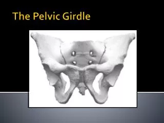
Pelvic girdle. Attaches lower limbs to the spine Supports visceral organs Attaches to the axial skeleton by strong ligaments Acetabulum is a deep cup that holds the head of the femur. Pelvic girdle. Consists of paired hip bones ( coxal bones) Hip bones unite anteriorly with each other


Pelvic girdle
E N D
Presentation Transcript
Pelvic girdle • Attaches lower limbs to the spine • Supports visceral organs • Attaches to the axial skeleton by strong ligaments • Acetabulum is a deep cup that holds the head of the femur
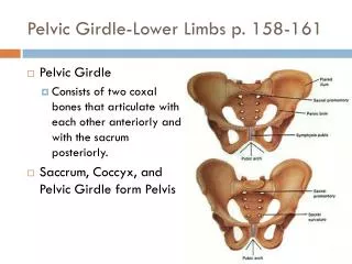
Pelvic girdle • Consists of paired hip bones (coxal bones) • Hip bones unite anteriorly with each other • Articulates posteriorly with the sacrum
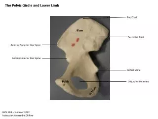
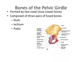
Hip bones • Consist of three separate bones in childhood • Ilium, ischium, and pubis • Bones fuse – retain separate names to regions of the coxal bones • The triradiatecartilage • Acetabulum • A deep hemispherical socket on lateral pelvic surface
ilium • The ilium is a large flaring bone that forms the superior region of the coxal. • It consists of a body and superior wing like portion called the ala • The broad posterolateral surface is called the gluteal surface • The auricular surface articulates with the sacrum (sacroiliac joint) • Major markings include the iliac crests, four spines, gretaer sciatic notch,iliacfossa,arcuate line and the pelvic
Ischium • Forms posteroinferior region of the coxal bone • Anteriorly – joins the pubis • Ischialtuberosities • Are the strongest part of the hip bone
Pubis • Forms the anterior region of the coxal bone • An angulated bone • Lies horizontally in anatomical position • Pubic symphysis (fribrocartilage)
True and False Pelves • Bony pelvis is divided into two regions • False (greater) pelvis – bounded by alae of the iliac bones • True (lesser) pelvis – inferior to pelvic brim • Forms a bowl containing the pelvic organs
Comparison of Male and Female Pelvic Structure • Female pelvis • –Tilted forward, adapted for childbearing • –True pelvis defines birth canal • –Cavity of the true pelvis is broad, shallow, and has greater capacity
Male pelvis • –Tilted less forward • –Adapted for support of heavier male build and stronger muscles • –Cavity of true pelvis is narrow and deep
Thigh • The region of the lower limb between the hip and the knee • Femur – the single bone of the thigh • Longest and strongest bone of the body • Ball-shaped head articulates with the acetabulum
Patella • Triangular sesamoid bone • Imbedded in the tendon that secures the quadriceps muscles • Protects the knee anteriorly • Improves leverage of the thigh muscles across the knee
Leg • Refers to the region of the lower limb between the knee and the ankle • The leg is fixed in permanent pronation • Composed of the tibia and fibula • Tibia – more massive medial bone of the leg • Receives weight of the body from the femur • Fibula – stick-like lateral bone of the leg • Interosseous membrane- Connects the tibia and fibula
tibia • Has 2 condyles- medial and lateral • Intercondylar eminence • Shaft has 3 surfaces- medial, lateral and posterior • Anterior border is most prominent and also called the shin or shin bone • Extends distally to form the medial malleolus • Fibular notch • Posterior surface has a soleal line • Nutrient foramen
fibula • Lies posteriolateral • Leg is fixed in permanent pronation • Distal end ends in lateral malleolus • Shaft has 3 borders (anterior, posterior and interosseous) and 3 surfaces (medial, posterior and lateral)
The Foot • Foot is composed of • Tarsus, metatarsus, and the phalanges • Important functions • Supports body weight • Acts as a lever to propel body forward when walking • Segmentation makes foot pliable and adapted to uneven ground
Tarsus • Makes up the posterior half of the foot • Contains seven (7)bones called tarsals • Talus, calcaneous, cuboid, navicular, 3 cuneiforms • Body weight is primarily borne by the talus and calcaneus
Metatarsus • Consists of five small long bones called metatarsals • Numbered 1–5 beginning with the hallux (great toe) • First metatarsal supports body weight
Phalanges of the Toes • 14 phalanges of the toes • Smaller and less nimble than those of the fingers • Structure and arrangement are similar to phalanges of fingers • Except for the great toe, each toe has three phalanges • Proximal, middle, and distal
fractures of the femur • Mostly age and sex related (elderly females <60) due osteoporosis • Most common site is the neck • Proximal femoral fractures • Transcervical fracture-femoral neck (avascular necrosis occurs due to retinacular arteries that are cut off from the medial circumflex femoral artery) • Inter trochanteric fracture • Femoral shaft fracture • spiral fracture (leads to foreshortening) • Distal femoral fractures • Fracture of femoral condyles- popliteal artery runs on the posterior surface
Tibial and fibula fracture • The tibial shaft is narrowest at the junction of its middle third and inferior thirds and most frequent site of fracture. • Poor blood supply • nutrient artery (nutrient canal posteriorly) • Types • Compound fracture (with external bleeding) • Diagonal fracture with shortening • Transverse fractures (with fibular intact) • March (stress) fracture
Fibula neck fracture • Direct trauma as nerve passes superficially around neck of fibula • footdrop and loss of eversion • May cause sensory loss over lateral leg and dorsum of foot
Fracture of Lateral malleolus Fibular malleolar fracture effect-excessive inversion of foot Common in soccer and football players
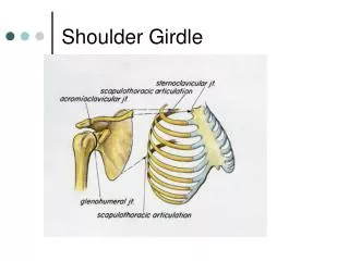
HIP JOINT, KNEE JOINT and ANKLE JOINT • Type • Articulation • Capsule • Ligaments • Movements • Blood Supply • Nerve Supply
Hip Joint • The hip joint forms the connection between the lower limb and the pelvic girdle
HIP JOINT • TYPE: BOLL & SOCKET TYPE • ARTICULATIONS : Cup shaped acetabulum & Hemi spherical head of femur Acetebular surface is horseshoe shaped Cavity is deepended by – fibro cartilagenous rim called “ Acetabular labrum”
5 IN NO. • ILIO-FEMORAL LIGAMENT: • - Strong, inverted “y” shaped Lig. • - Base is above – from AIIS • - 2 limbs are below from – • upper & lower parts of Inter – • trochanteric line • 2. PUBO - FEMORAL LIGAMENT: • - Triangular in shape • Base – superior ramusof pubis • Apex – Lower part of Inter trochanteric line • Limits – extension & abduction LIGAMENTS


![Download Book [PDF] Sacroiliac Pain: Understanding the Pelvic Girdle Musculoskeletal Method](https://cdn7.slideserve.com/12525915/slide1-dt.jpg)