Download
1 / 47
530 likes | 959 Vues
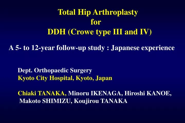
Total Hip Arthroplasty for DDH (Crowe type III and IV). A 5- to 12-year follow-up study : Japanese experience. Dept. Orthopaedic Surgery Kyoto City Hospital, Kyoto, Japan Chiaki TANAKA, Minoru IKENAGA, Hiroshi KANOE, Makoto SHIMIZU, Koujirou TANAKA. Purpose.

E N D
Total Hip Arthroplasty for DDH (Crowe type III and IV) A 5- to 12-year follow-up study : Japanese experience Dept. Orthopaedic Surgery Kyoto City Hospital, Kyoto, Japan Chiaki TANAKA, Minoru IKENAGA, Hiroshi KANOE, Makoto SHIMIZU, Koujirou TANAKA
Purpose After my study in Cochin Hospital, I began to operate on the DDH patients in Japan. Especially, THA for DDH (Crowe type III and IV) patients is a technically demanding operation. We report our 5- to 12- year clinical results and technical problems in THA for difficult DDH patients.
The most difficult case in my experience Case K.K. 62yr-old F : 134.5 cm 54.3 kg Crowe IV 88/10/22 89/3/8 88/11/10 93/2/2
Case K.K. 62yr-old F : Crowe IV 93/12/24 94/9/29 94/1/14 95/4/6
Case K.K. 62yr-old F : Crowe IV 95/4/6 04/10/08 9yr 5mo
PATIENTS - 1 32 hips (27 women, 1 man) Average age at operation 58.5 y.o. ( 44-78 ) Follow-up period 8 yr. 3 mo. ( 5-12 y ) Body weight 49.3 kg ( 35.7 – 67.0 ) Height 148.5cm ( 134.5 – 167.2)
PATIENTS - 2 Crowe type III 14 IV 18 Previous Operations none 25 femoral osteotomy 7 pelvic osteotomy 1
RECONSTRUCTIVE METHOD-1 Lateral transtrochanteric approach 32 Total capsulectomy 32 Muscle release 0 Bone grafting acetabular 32 femoral 1 THA device Charnley LFA 8 Kyocera PHS 19 CMK 5 32
RECONSTRUNTIVE METHOD-2 Cup diameter 37 1 38 6 38.5 3 40 15 42 2 44 3 46 2 Fixation of greater trochanter Ortron wire27 Dall-Miles Cable Grip3 Titanium wire2
Clinical and Radiographical Evaluation Japanese Orthopaedic Association (JOA) Hip Score System Pain 40, ROM 20, Walking ability 20, ADL 20 Radiolucency : DeLee – Charnley zone Migration > 3mm or > 3degrees Position of hip center : distance from teardrop Bone graft coverage : % of the cup Bone grafts : union, resorption, collapse
RESULTS Revision : (acetabular loosening) 1 Reoperation : trochanter reattach 1 abductor advancement 1 32 Complications Dislocation 0 Trochanteric nonunion 4 Infection 0 Nerve palsy 0 32
JOA Hip Score Preop Last FU Pain 16.7 39.1 ROM 9.7 16.1 Walk 5.0 12.1 ADL 8.5 14.6 Total 39.8 81.9
Radiographic Evaluation - 1 Migration (Cup) 1 (Rev) Radiolucency Acetabular none 25 partial 6 (osteolysis 1) 32 Acetab. Loosening 1 / 32 ( 6.3%) PE Wear 2mm< 1
Radiographic Evaluation - 2 Stem sinking 0 Radiolucency partial 1 Osteolysis severe 2 mild 2
Radiographic Evaluation - 3 Rotational hip center horizontal distance av. 29.7mm vertical distance av. 22.4mm Bone graft coverage B / A av. 38% ( 24 ~ 54 ) 50%< 5 hips collapse 1 hip (Rev. at 18 mo) B A
Survivorship Endpoint : Revision 10 years Acetabular component 96.9% Femoral component 100%
THA for DDH Mackenzie JR 1996 59 hips (II:22,III:18,IV:19) Surv(Rev) 85% at 15y Surv(Rad.loose) 68% Numair J 1997 46 hips (IV) Surv(Rev) 68% at 15y Shinar AA 1977 70 hips Rev 36% Rev+Rad loose 60% at 16.5y Bobak P 2000 45 hips (I:4,II:17,III:13,IV:11) Rev 0% Rad loose 12% at 11y (10-15) Kerboull M 2001 118 hips (IV) Surv 78% at 20 y Kobayashi S 2003 37 hips (II:16,III:17,IV:4) Rev 0% Rad Loose 0% at 19 y (10-26) Hartofilakidis G 2004 84 hips (high disl) Surv(Rev) 76.4% at 15 y
Case K.K. 60yr-old F : Crowe III Pre-op 2mo 10yr 12.5yr JOA score 83 p. Wear < 2mm Osteolysis
Case M.K. 50yr-old F : Crowe IV Pre-op 1 mo 11 yrs JOA score 92 p.
Case S.I. 55yr-old F : Crowe IV Pre-op 2mo 8mo 9mo 12yr JOA s. 78 p. Nonunion Gr.Tr. Titanium wire Nonunion of Gr.Tr. Troch. Rev.
Case T.H. 78yr-old F : Crowe III Preop 2 mo 7 yr 9 mo JOA score 94 p.
Case Y.S. 80yr-old F : Crowe IV Pre-op 2 mo 6 yr 3 mo JOA score 76 p.
Case M.I. 53yr-old F : Crowe IV Preop 9 y 10 mo 9 y 5 mo JOA score 81 p.
Case F.M. 57yr-old F : Crowe III Advancem. 1w Preop 8yr8mo 3w Abduction contracture 25 deg. Trendelenbourg (-) JOA score 74 p.
Problems and my solutions • Acetabulum • Small and shallow acet. thin walls • CT scan is useful • Difficult exposure of the true acetabulum • Joint capsule is the excellent guide • Bone grafting • Deficient superior and posterior wall • Preservation of ant. and post. ‘column horn’ • Small and atrophic femoral head • graft shaping method • option: harvesting the cortical bone • by shortening the femoral shaft
Case K.K. 62yr-old F : Crowe IV Small Acetabulum True acetabulum is the best position !!
Though the true acetabulum is the best position, the AP diameter is small. 40 mm
Problems and my solutions • Acetabulum • Small and shallow acet. thin walls • CT scan is useful • Difficult exposure of the true acetabulum • Joint capsule is the excellent guide ‘Asagao’ • Bone grafting • Deficient superior and posterior wall • Preservation of ant. and post. ‘column horn’ • Small and atrophic femoral head • graft shaping method • option: harvesting the cortical bone • by shortening the femoral shaft
Joint capsule : the best guide to obturator foramen It looks like a Morning Glory:‘Asagao’ in japanese Morning Glory : ‘Asagao’ Femur Greater Troch External obturator
Problems and my solutions • Acetabulum • Small and shallow acet. thin walls • CT scan is useful • Difficult exposure of the true acetabulum • Joint capsule is the excellent guide • Bone grafting • Deficient superior and posterior wall • Preservation of ant. and post. ‘column horn’ • Small and atrophic femoral head • graft shaping method • option: harvesting the cortical bone • by shortening the femoral shaft
Problems and my solutions • Acetabulum • Small and shallow acet. thin walls • CT scan is useful • Difficult exposure of the true acetabulum • Joint capsule is the excellent guide • Bone grafting • Deficient superior and posterior wall • Preservation of ant. and post. ‘Column Horn’ • Small and atrophic femoral head • graft shaping method • option: harvesting the cortical bone • by shortening the femoral shaft
Preservation of ant. and post. ‘Column Horn’ Case M.K. 50yr-old F : Crowe IV 3D-CT Image
Problems and my solutions • Acetabulum • Small and shallow acet. thin walls • CT scan is useful • Difficult exposure of the true acetabulum • Joint capsule is the excellent guide • Bone grafting • Deficient superior and posterior wall • Preservation of ant. and post. ‘column horn’ • Small and atrophic femoral head • graft shaping method • option: harvesting the cortical bone • by shortening the femoral shaft
Problems and my solutions • Acetabulum • Small and shallow acet. thin walls • CT scan is useful • Difficult exposure of the true acetabulum • Joint capsule is the excellent guide • Bone grafting • Deficient superior and posterior wall • Preservation of ant. and post. ‘column horn’ • Small and atrophic femoral head • graft shaping method • option: harvesting the cortical bone • by shortening the femoral shaft
Problems and my solutions 2) Femoral side Narrow canal, strong anteversion straight stem Respect the greater trochanteric bone bed when the femoral neck is cut stem design is very important !! Subtrochanteric shortening osteotomy is a useful technique when the reduction seems very difficult or when the femoral neck needs to be cut too much or in previously osteotomized cases
Preservation of Trochanter Bed * Kyocera PHS CMK
Problems and my solutions 2) Femoral side Narrow canal, strong anteversion straight stem Respect the greater trochanteric bone bed when the femoral neck is cut stem design is very important !! Subtrochantericshortening osteotomy is a useful technique when the reduction seems very difficult or when the femoral neck needs to be cut too much or in previously osteotomized cases
Extra-series Case T.S. 70yr-old F : Crowe IV Case S.K. 56yr-old F : Crowe IV
Problems and my solutions 3) Limb lengthening If the range of motion is good, lengthening is easy. If not, removal of the scar tissue is necessary. Principles of Prof. Kerboull Respect the periarticular muscles as possible. The best method to avoid nerve palsy !! Reduction mild flexion and adduction of the hip with mild flexion of the knee pushing the stem head directly into the cup Never pull the limb !!
Problems and my solutions 4) Trochanter fixation In severely contracted hips, lowering the greater trochanter is difficult. Detachment of gluteal muscle origin upwards from ilium Option : advancement of gluteal muscles through the iliac rest incision Fixation with stainless monofilament wires Attention to titanium wires and Dall-Miles cables !!
Lowering of Greator Trochanter Advancement Detachment upwards from ilium M. Kerboull EMC R.C. Kingsley JBJS
Problems and my solutions 4) Trochanter fixation In severely contracted hips, lowering the greater trochanter is difficult. Detachment of gluteal muscle origin from ilium upward direction from inside Option : advancement of gluteal muscles through the iliac rest incision Fixation with stainless monofilament wires Attention to titanium wires and Dall-Miles cables !!
Fixation of Greator Trochanter Attention to Titanium wires and Dall-Miles Cables Titanium wire Dall-Miles Cable Grip
Conclusions THA for DDH (Crowe type III and IV) patients is a technically demanding operation. 5- to 12- year clinical results of our series were satisfactory. Main techinical problems are reconstruction of very small dysplastic acetabuli and solid fixation of greater trochanter.