Herpes viruses
Herpes viruses. Introduction. Leading cause of human viral disease, second only to influenza & cold viruses. Capable of causing overt disease or remaining silent for many years only to be reactivated, e.g. shingles

Herpes viruses
E N D
Presentation Transcript
Introduction • Leading cause of human viral disease, second only to influenza & cold viruses. • Capable of causing overt disease or remaining silent for many years only to be reactivated, e.g. shingles • Derived from the Greek word herpeinmeans tocreep……reflects the creeping or spreading nature of the skin lesions caused by herpes virus.
Structure Large, enveloped DNA viruses Icosahedralcapsids DNA replicates in the nucleus of host cells They form Cowdrytpye A intranuclear inclusion bodies
This family includes 8 human pathogens: • Herpes simplex viruses (HSV) types 1 &2 • Varicella Zoster virus (VZV) • Cytomegalo virus (CMV) • Epstein-Barr virus (EBV) • Human Herpes viruses (HHV) types 6,7&8
Types of Herpesviruses Dr Ekta Chourasia, Microbiology
General Characteristics • Most infections are asymptomatic • They remain latent in the host after recovery from primary infection • All herpes viruses cause recurrent infections by reactivation of viral replication • In tissue culture they produce: • Intracellular inclusion bodies • Cytopathic effect: ballooning and rounding of infected cells followed by cell death • Some are oncogenic: HSV-2, EBV, and HHV-8 • They can cause severe disease in immunocompromised hosts and neonates
Herpes Simplex Viruses • Extremely wide spread in human population • Establish latency in nerve cells • Reactivation is common • There are two distinct types of HSVs: type 1 &type 2 • Structurally & morphologically indistinguishable • Exhibit sequence gene homology with serological cross-reaction • Can be distinguished by restriction enzyme analysis of viral DNA and mode of transmission
Pathogenesis of HSV 1 &2 Sensory ganglia replication Initial infection site Migration through Neuron reactivation Reactivation is through stress stimuli such as UV light, fever, hormonal changes, surgical trauma to the neuron latency Antibodies do not prvent reactivation HSV-1: trigeminal ganglia HSV-2: sacral ganglia
Diseases caused by HSV-1 1- Oropharyngeal infections: • Acute gingivostomatitis: occurs in early childhood • Fever, • painful vesicular lesions ; on gums, lips & oral mucosa, these vesicles may rupture leaving a red based ulcer which • may be 2ry infected with candidaalbicans forming white coat • Herpes labialis (fever blisters or cold sores) • Milder recurrent form • Crops of vesicles at the mucocutaneous junction of lips or nose
Herpes Labialis Acute herpetic ginfivostomatitis
2- Herpetic Keratoconjunctivitis: • Corneal ulcers and lesions of conjunctival epithelium • Recurrence takes the appearance of dendritic ulcer or vesicles on the eye lids • Recurrent keratitis may lead to permanent scarring ending with blindness
Keratitis Vesicles around eye lid
3- Encephalitis • Rare • Involves temporal lobe with high mortality 4- Herpetic Whitlow: • Fingers herpes infection • In health care workers e.g. dentists & nurses 5- Eczema Herpeticum: • Involves known eczematous areas with bacterial superinfection 6- Disseminated infection: Fatal esophagitis or pneumonia in immunocompromised patients
Herpetic Withlow Eczema Herpeticum
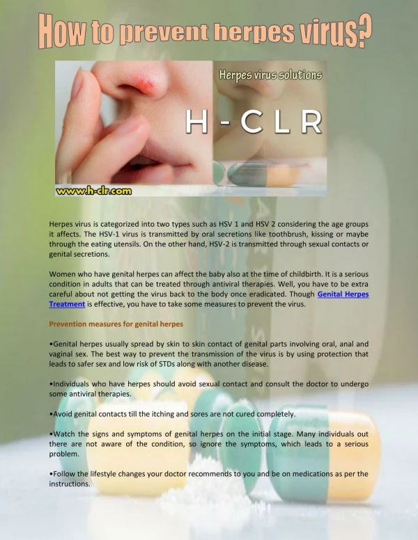
Diseases caused by HSV-2 1- Genital herpes: • Sexually transmitted • Vesiculoulcerative lesions of penis in males and cervix, vulva, vagina, & perineum of females 2- aseptic meningitis: Self limited 3- Neonatal Herpes: Acquired in utero,during, or after birth Severe in the newborn so, pregnant females with recurrent herpes should deliver by CS
Laboratory Diagnosis • Specimen: Vesicular fluid- Corneal scrapping 1- Direct Virus Demonstration: a) L/M: • Tzanck smear – from the base of vesicles, 1% aq. soln. of toluidine blue ‘O’ shows multinucleated giant cells with faceted nuclei & homogenously stained ‘ground glass’ chromatin (Tzanck cells) • Giemsa stained smear – intranuclearCowdry type A inclusion bodies
Tzanck smear intranuclearCowdry type A inclusion bodies
B) Direct Immunofluorescence: • Cell scrapings from lesions are stained with monoclonal antibodies conjugated with a fluorescence dye. Viral inclusion bodies appear in UV microscope as a bright green intranuclear particles • C) PCR: for detection of viral DNA in CSF
2- Viral Isolation: tissue culture: human diploid fibroblasts, human amnion, human embryonic kidney: CPC (syncytium formation) seen in 24-48 hrs. • 3) Serology: useful in the diagnosis of primary infection, Ab (IgM) detection by ELISA, NT or CFT.
Treatment • Inhibits viral DNA polymerase enzyme • topical, oral, or IV Acyclovir Doesn’t affect latency
Varicella –Zoster Virus (VZV) Causes 2 major diseases • Varicella (chicken pox): primary infection usually in childhood • Zoster ( shingles): reactivation of an earlier varicella
Varicella (Chicken Pox) • Mild, highly contagious disease chiefly affecting children • Mode of transmission: - airborne droplets and direct contact from varicella patients - Vesicular fluid of Zoster patients can be the source of Varicella in susceptible children
Pathogenesis: • VZV infects the mucosa of the upper respiratory tract • Multiplies in the regional LNs • Primary viremia and spread to liver and spleen • Secondary viremia follows with viral spread to the skin • Typical rash occurs • VZV remains latent in the dorsal root ganglia for life
Clinical Picture: • Incubation period: 10-21 days • Symptoms: mild fever & rash • Rash: first appears on the trunk, then face and limbs • Flat macules become papules then vesicles • Followed by crust formation • The crust is often shed off and heals without scarring • Cropping is a characteristic feature of varicella rash: fresh vesicles appear in crops, so that all stages of macules, papules, vesicles & crusts are seen at the same time • More severe in adults
Complications • 1- pneumonia especially in adults, may be fatal • 2- rarely: fulminant encephalitis, which may be a manifestation of Reye’s syndrome that occurs as a consequence of salicylates intake during infection
Congenital Varicella Syndrome & Neonatal Varicella • Primary maternal infection during the 1st trimester may lead to congenital varicella syndrome ( serious & fatal): skin lesions, hypoplasia of limbs, chorioretinitis & CNS defects • Primary maternal infection near the time of birth can lead to widely disseminated infection in the new born with mortality rate of 35% • If rash began a week or more before delivery, maternal Abs transferred via placenta – baby gets the infection but escapes clinical disease
Zoster (shingles) • Sporadic disease in adults or immunocompromised patients • Results from reactivation of latent VZV • Rash similar to varicella but limited to a nerve distribution to the skin innervated by a dorsal root ganglion (dermatom)
Complications: • If affecting the eye via trigeminal nerve: keratitis, conjunctivitis & iritis • It can affect the brain via the cranial nerve leading to Bell’s palsy • Post Herpetic neuralgia: Very painful, Likely due to nerve damage from zoster outbreak, Lasts for months after zoster resolves & Does not respond to antiviral treatment
Laboratory Diagnosis • Specimen: Vesicular lesions smears 1- Direct Virus Demonstration: a) L/M: Tzanck smear – from the base of vesicles, 1% aq. soln. of toluidine blue ‘O’ shows multinucleated giant cells with faceted nuclei & homogenously stained ‘ground glass’ chromatin (Tzanck cells)
B) Direct Immunofluorescence: • Cell scrapings from lesions are stained with monoclonal antibodies conjugated with a fluorescence dye. Viral inclusion bodies appear in UV microscope as a bright green intranuclear particles • C) PCR: for detection of viral DNA 2- serology: • Specific VZV Abs using CFT, Nt, or ELISA
Treatment • Inhibits viral DNA polymerase enzyme • Used in immunocompromised patients with chicken pox, zoster, or varicella complicated with pneumonia, keratitis, & neonatal Varicella Acyclovir Doesn’t affect latency
Prevention 1- Active immunization 2- Passive immunization Varicellazoster immunoglobulins (VZIG) Given to: Immunocompromised children exposed to infection Mothers infected near term(before delivery) and their infants ( immediately after delivery) • Live attenuated varicella vaccine Single dose, age: 1-12 yrs