Download
1 / 52
520 likes | 665 Vues
Lab Chapter 27 Functional Anatomy of the Endocrine Glands. Major Endocrine Organs. Figure 16.1. Hormones. Hormones – chemical substances secreted by cells into the extracellular fluids Regulate the metabolic function of other cells

E N D
Lab Chapter 27 Functional Anatomy of the Endocrine Glands
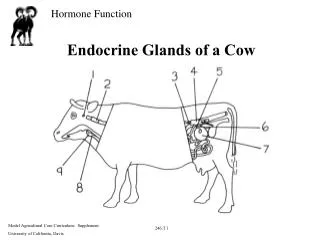
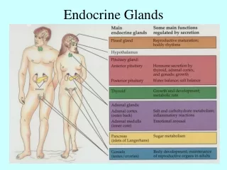
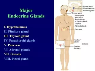
Major Endocrine Organs Figure 16.1
Hormones • Hormones – chemical substances secreted by cells into the extracellular fluids • Regulate the metabolic function of other cells • The precise response depends on the type of the target cell
Target Cell Specificity • Hormones circulate to all tissues but only activate cells referred to as target cells • Target cells must have specific receptors to which the hormone binds
Gross Anatomy -Hypophysis • Pituitary gland – two-lobed organ that secretes nine major hormones • Neurohypophysis – posterior lobe (neural tissue) and the infundibulum • Receives, stores, and releases hormones from the hypothalamus • Adenohypophysis – anterior lobe, made up of glandular tissue • Synthesizes and secretes a number of hormones
Pituitary (Hypophysis) Figure 16.6
Pituitary-Anterior Lobe • There is a vascular connection, the hypophyseal portal system, consisting of: • The primary capillary plexus • The hypophyseal portal veins • The secondary capillary plexus
Adenophypophyseal Hormones • The six hormones of the adenohypophysis: • Abbreviated as GH, TSH, ACTH, FSH, LH, and PRL
Activity of the Adenophypophysis • The hypothalamus sends a chemical stimulus to the anterior pituitary • Releasing hormones stimulate the synthesis and release of hormones • Inhibiting hormones shut off the synthesis and release of hormones
Activity of the Adenophypophysis • The tropic hormones • Stimulates an endocrine gland instead of a target organ. They are: • Thyroid-stimulating hormone (TSH) • Adrenocorticotropic hormone (ACTH) • Follicle-stimulating hormone (FSH) • Luteinizing hormone (LH)
Thyroid Stimulating Hormone (Thyrotropin) • Stimulates the normal development and secretory activity of the thyroid • Triggered by hypothalamic peptide thyrotropin-releasing hormone (TRH)
Adrenocorticotropic Hormone (Corticotropin) • Stimulates the adrenal cortex to release corticosteroids • Triggered by hypothalamic corticotropin-releasing hormone (CRH) in a daily rhythm
Gonadotropins • Gonadotropins – follicle-stimulating hormone (FSH) and luteinizing hormone (LH) • Regulate the function of the ovaries and testes • FSH stimulates gamete (egg or sperm) production • Triggered by the hypothalamic gonadotropin-releasing hormone (GnRH)
Functions of Gonadotropins • In females • LH promotes synthesis and release of estrogens and progesterone • In males • LH stimulates synthesis of testosterone by the testes
Adenohypophysis- Growth Hormone (GH) • Stimulate most cells, but target bone and skeletal muscle • Promote protein synthesis and encourage the use of fats for fuel • Dwarfism, gigantism, acromegaly
Prolactin (PRL) • In females, stimulates milk production by the breasts • Triggered by the hypothalamic prolactin-releasing hormone (PRH)
Pituitary - Posterior Lobe • The posterior lobe is a downgrowth of hypothalamic neural tissue • Has a neural connection with the hypothalamus (hypothalamic-hypophyseal tract) • Nuclei of the hypothalamus synthesize oxytocin and antidiuretic hormone (ADH) • These hormones are transported to the posterior pituitary
The Posterior Pituitary and Hypothalamic Hormones • ADH influences water balance • Produced by the supraoptic nuclei of the hypothalamus • Oxytocin stimulates smooth muscle contraction in breasts and uterus • Produced by the paraventricular nuclei of the hypothalamus
Thyroid Gland • Consists of two lateral lobes connected by the isthmus
Thyroid Gland Figure 16.8
Thyroid Hormone • Consists of two related iodine-containing compounds • T4 – thyroxine; has two tyrosine molecules plus four bound iodine atoms • T3 – triiodothyronine; has two tyrosines with three bound iodine atoms
Effects of Thyroid Hormone • TH is concerned with: • Increasing metabolic rate • Heat production • Cellular oxidation • Hyperthyroidism • Hypothyroidism
Calcitonin • Produced by the parafollicular, or C, cells • Lowers blood calcium levels • Antagonist to parathyroid hormone (PTH)
Parathyroid Glands • Tiny glands embedded in the posterior aspect of the thyroid • PTH (parathormone) increases the level of calcium in the blood
Parathyroid Glands Figure 16.11
Effects of Parathyroid Hormone • PTH release increases Ca2+ in the blood as it: • Stimulates osteoclasts to digest bone matrix • Enhances the reabsorption of Ca2+ and the secretion of phosphate by the kidneys • Increases absorption of Ca2+ by intestinal mucosal • Hyperparathyroidism • Hypoparathyroidism
Effects of Parathyroid Hormone Figure 16.12
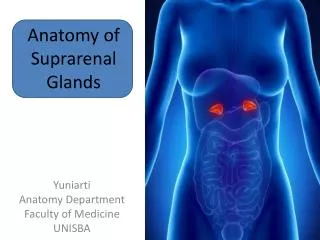
Adrenal (Suprarenal) Glands • Adrenal glands – paired, pyramid-shaped organs atop the kidneys • Structurally and functionally, they are two glands in one • Adrenal medulla – neural tissue that acts as part of the SNS • Adrenal cortex – glandular tissue derived from embryonic mesoderm
Adrenal Cortex • Synthesizes and releases steroid hormones called corticosteroids • Different corticosteroids are produced in each of the three layers • Zona glomerulosa – mineralocorticoids (chiefly aldosterone) • Zona fasciculata – glucocorticoids (chiefly cortisol) • Zona reticularis – gonadocorticoids (chiefly androgens)
Mineralocorticoids • Water balance in extracellular fluids • Electrolyte balance in extracellular fluids • Aldosterone – most important mineralocorticoid • Stimulates reabsorption of Na+ by the kidneys
Glucocorticoids (Cortisol) • Help the body resist chronic stress by: • Keeping blood sugar levels relatively constant • Cortisone, hydrocortisone, corticosterone: • Rises in blood glucose, fatty acids, and amino acids
Gonadocorticoids (Sex Hormones) • Most gonadocorticoids secreted are androgens (male sex hormones), and the most important one is testosterone • Androgens contribute to: • The onset of puberty • The appearance of secondary sex characteristics • Sex drive in females
Adrenal Medulla • Secrete epinephrine and norepinephrine • Secretion of these hormones causes: • Blood glucose levels to rise • Blood vessels to constrict • The heart to beat faster • Blood to be diverted to the brain, heart, and skeletal muscle
Pancreas • A triangular gland, which has both exocrine and endocrine cells, located behind the stomach • Acinar cells produce an enzyme-rich juice used for digestion (exocrine product) • Pancreatic islets (islets of Langerhans) produce hormones (endocrine products) • The islets contain two major cell types: • Alpha () cells that produce glucagon • Beta () cells that produce insulin
Insulin and Glucagon • Glucagon major target is the liver, where it promotes: • Release of glucose to the blood from liver cells • Insulin: • Lowers blood glucose levels • Enhances transport of glucose into body cells • Counters metabolic activity that would enhance blood glucose levels
Gonads: Female • Paired ovaries in the abdominopelvic cavity produce estrogens and progesterone and the ova • Estrogen: • Appearance of secondary sexual characteristics • Breast development and cyclic changes in the uterine mucosa
Gonads: Female • Progesterone: • Cycling changes of the uterine lining • Keeps pregnancy • Prepares breast for lactation
Gonads: Male • Testes located in an extra-abdominal sac (scrotum) produce testosterone • Testosterone: • Initiates maturation of male reproductive organs • Causes appearance of secondary sexual characteristics and sex drive • Is necessary for sperm production • Maintains sex organs in their functional state
Pineal Gland • Small gland hanging from the roof of the third ventricle of the brain • Secretory product is melatonin • Melatonin is involved with: • Day/night cycles • Physiological processes that show rhythmic variations (body temperature, sleep, appetite)
Thymus • Lobulated gland located deep to the sternum • Major hormonal products are thymopoietins and thymosins • These hormones are essential for the development of the T lymphocytes (T cells) of the immune system
Microscopic anatomy of the glands • Thyroid gland • Follicles – simple squamous or cuboidal cells • Colloid – contain thyroglobulin, T3 and T4 • Parafollicular cells (C cells) • Between follicles • Secrete calcitonin
Microscopic anatomy of the glands • Parathyroid • Fibrous capsule surrounds the organ • Chief cell • Small cells, round nuclei, arranged in clusters, secret PTH • Oxyphil cells • Lager than chief cells • Scattered • Unknown function
Microscopic anatomy of the glands • Pancreas • Exocrine acini • Darker cells • Endocrine with Islets of Langehans • Beta cells – located at the center and secrete insulin • Alpha cells – located at the periphery and secrete glucagon
Microscopic anatomy of the glands • Anterior Pituitary gland • Chromophils • Acidophils- reddish-brown. GH and PRL • Basophils – deep-blue. Tropic hormones • Chromophobes • Do not take collor (Pale) • Controversial role
Microscopic anatomy of the glands • Posterior pituitary gland • Nerve fibers • Pituicytes (glial cells)
Microscopic anatomy of the glands • Adrenal gland- cortex • Zona glomerulosa • outermost, spherical clusters of cells, secrete mineralocorticoids • Zona fasciculate • Next layer, cells arranged in parallel cords, secrete glucocorticoids • Zona reticularis • Inneremost, cells arranged in branches, produces mainly sex hormones
Microscopic anatomy of the glands • Adrenal gland – medulla • The core of the gland • Modified neurons • Secrete E and NE
Microscopic anatomy of the glands • Ovary • Vesicular follicle • Ovum