Download
1 / 44
440 likes | 642 Vues
Adult Health Nursing II Block 7.0. Topic: Cardiovascular Nursing, & EKG Monitoring, part 3 Module: 2.4. Cardiovascular--- EKG’s / Cardiac Monitoring. Dynamic Presentation . Static Presentation. Lead II. Part III. Digitalis pupurea (Foxglove). Key Terms.

E N D
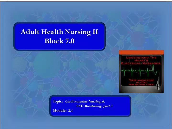
Adult Health Nursing II Block 7.0 Topic: Cardiovascular Nursing, & EKG Monitoring, part 3 Module: 2.4
Cardiovascular--- EKG’s / Cardiac Monitoring Dynamic Presentation Static Presentation Lead II Part III Block 7.0 Module 2.4 Digitalis pupurea (Foxglove)
Key Terms • Arrhythmia & Dysrhythmia • Electrical Cardioversion • Defibrillation • The “Names” of all of the rhythms & dysrhythmias • atropine • amiodarone • lidocaine (Xylocaine ®) • adenosine (Adenocard ®) • dopamine • epinephrine • nitroprusside (Nipride ®) Block 7.0 Module 2.4
Physical Assessment: S/S of Decreased Cardiac Output Block 7.0 Module 2.4
General Method…. • General Impression • Rate= ________ • Rhythm =_______ • P Waves =_______ • PRI=_______ • QRS = _______ • Fast, “tight” QRS’s, fairly regular, no “FLB’s” • Rate= 120’s • Rhythm = Regular • P Waves = Present, upright, uniform, 1:1 ratio w/QRS’s, (precede QRS) • PRI = 0.16 seconds, = throughout strip • QRS = 0.08 seconds Block 7.0 Module 2.4
General Impression • Rate=___________ • Rhythm=_________ • P Waves: ________ • PRI= __________ • QRS = __________ • Medium rate, funny-looking P’s, no FLB’s • 100’s • Regular • Present, upright, ~, biphasic, inverted, or “s”-shaped, 1:1 w / QRS’s • 0.10 seconds • 0.08 seconds Block 7.0 Module 2.4
Normal Sinus Rhythm RATE: 60-100 RHYTHM: Regular P Waves: Upright, uniform (~), 1:1 with QRS Complexes PR Interval: 0.12 – 0.20 seconds QRS: < 0.12 sec, ~ Block 7.0 Module 2.4
Sinus Bradycardia RATE: < 60 RHYTHM: Regular P Waves: Upright, uniform, 1:1 with QRS Complexes PR Interval: 0.12 – 0.2 seconds, uniform QRS: < 0.12 sec, ~ Discussion: May be benign; Treatment Atropine IVP for Symptomatic Bradycardia Block 7.0 Module 2.4
Sinus Tachycardia RATE: 100 -150 RHYTHM: Regular P Waves: Upright, uniform (~), 1:1 with QRS Complexes PR Interval: 0.12 – 0.20 seconds, uniform (~) QRS: < 0.12 sec, ~ Discussion: Etiology? Block 7.0 Module 2.4
Atrial Flutter RATE: Variable; RHYTHM: Regular or Irregular P Waves: Absent; Instead, heave F – Waves, or Flutter Waves PR Interval: N/A QRS: < 0.12 sec Discussion: Rhythm may be regular or irregular, depending on ventricular response. Typically expressed as a “ratio,”, e.g., the above would be described as “Atrial flutter with a 3:1 block.” VERY COMMON AFTER ANY TYPE OF CARDIAC SURGERY; FREQUENTLY PROGRESSES TO ATRIAL FIBRILLATION; MAY “BOUNCE BACK & FORTH” “A-Fib-Flutter” or “A-Flutter-Fib” Block 7.0 Module 2.4
Atrial Fibrillation RATE: Variable; Rate may indicate effect on Cardiac Output (Loss of “Atrial Kick,” ~ 20 % C.O.) RHYTHM: Irregular P Waves: Absent PR Interval: N/A QRS: < 0.12 sec Discussion: -Most common dysrhythmia -Classified as “AF with controlled ventricular response,” “AF with rapid ventricular response,” “Uncontrolled AF.” -Treatment: Digoxin; cardioversion -Embolus Role in CVA & PE CHF Block 7.0 Module 2.4
DISCUSSION: Atrial Fibrillation Untreated or “uncontrolled Atrial fibrillation “ is a rapid and irregular heart arrhythmia, caused by chaotic electrical impulses in the atria of the heart (the two upper chambers). (Loss of “Atrial Kick,” i.e., ~ 20% of Cardiac Output) In anatomical terms, the AV node and the ventricles (the two lower chambers) are therefore bombarded with frequent, irregular electrical impulses. As a result, the heart rate becomes fast and irregular, and the normal coordination between the atria and the ventricles is lost. There are several types, depending on how long the AF lasts. When atrial fibrillation is always present, it is referred to as chronic atrial fibrillation. When the arrhythmia is usually present, such that episodes of normal rhythm are infrequent or short-lived, it is referred to as persistent atrial fibrillation. When a normal heart rhythm is usually present but occasional episodes of the arrhythmia occur, the patient is said to have paroxysmal atrial fibrillation. Block 7.0 Module 2.4
Supraventricular Tachycardia RATE: 151 – 220+ RHYTHM: Regular P Waves: Absent (buried in QRS) PR Interval: N/A QRS: < 0.12 sec Discussion: C.O. is decreased due to lack of ventricular filling time. Treatment: Vagal Maneuvers (Carotid Massage) Adenosine IVP Cardioversion REMEMBER:“Narrow-Complex Tachycardia” Block 7.0 Module 2.4
Discussion: Supraventricular tachycardias (SVT--PSVT) The SVTs are generally benign (that is, non-life-threatening) tachycardias that either arise in the atria (that is, “supra” the ventricles), or involve the atria in the mechanism of the tachycardia. Many SVTs are due to extra, abnormal electrical connections between the atria and the ventricles. Individuals with SVT are often born with these extra pathways. The existence of such extra pathways (often called “bypass tracts”) allow the formation of “reentrant” arrhythmias, in which an electrical impulse is established that spins continuously between the atria and the ventricles, thus causing one form of SVT. Wolff-Parkinson-White (WPW) syndrome is a common example, but there are several other varieties of bypass tracts that can cause episodes of SVT. Block 7.0 Module 2.4
Wolf-Parkinson White Syndrome "WPW is a form of supraventricular tachycardia (fast heart rate originating above the ventricles). Block 7.0 Module 2.4
WPW…. "WPW is a form of supraventricular tachycardia (fast heart rate originating above the ventricles). When you have WPW, along with your normal conduction pathway, you have extra pathways called accessory pathways. They look like normal heart muscle, but they may: --conduct impulses faster than normal --conduct impulses in both directions The impulses travel through the extra pathway (short cut) as well as the normal AV-HIS Purkinje system. The impulses can travel around the heart very quickly, in a circular pattern, causing the heart to beat unusually fast. This is called re-entry tachycardia. Re-entry arrythmias occur in about 50 percent of people with WPW; some may have atrial fibrillation (a common irregular heart rhythm distinguished by disorganized, rapid, and irregular heart rhythm). The greatest concern for people with WPW is the possibility of having atrial fibrillation with a fast ventricular response that worsens to ventricular fibrillation, a life-threatening arrhythmia,. Block 7.0 Module 2.4
Junctional Rhythms A.K.A. “AV Junctional Rhythms” But, this rate can be widely variable! RATE: 40-60 RHYTHM: Regular P Waves: Inverted, absent, or retrograde (after QRS) PR Interval: < 0.12 sec, or absent QRS: < 0.12 sec, ~ Discussion: Rate > 60= “Accelerated Junctional Rhythm;” Greater than 100= “Junctional Tachycardia” Block 7.0 Module 2.4
Junctional Tachycardia • Rate: 101 • Rhythm: Regular • P Wave: inverted, = , ~, 1:1 w/QRS’s • PRI = 0.08-0.10 sec, ~ • QRS = 0.06- 0.08 sec, ~ Block 7.0 Module 2.4
AV Blocks The specialized conduction system is responsible for transmitting the heart’s electrical impulses from the atria to the ventricles. Disease in the AV node, bundle of His, or the bundle branches can lead to a condition called “heart block.” Heart block occurs when the electrical impulses in the atria are stopped from reaching the ventricles. The heart rate can reach dangerously low levels when heart block is present. A permanent pacemaker, however, takes care of the problem. Block 7.0 Module 2.4
1st Degree AV Block RATE: Variable RHYTHM: Regular P Waves: Present, upright, uniform, 1:1 ratio with QRS PR Interval: Uniform, > 0.20 sec QRS: < 0.12 sec Discussion: usually benign The above rhythm would be described as: “Sinus Rhythm, 1st Degree AV Block, Rate=_______ Block 7.0 Module 2.4
Sinus Tachycardia, 1st Degree AV Block Block 7.0 Module 2.4
2nd Degree AV Block(Mobitz I --”Wenkebach”--and Mobitz II) RATE: Variable, usually slow RHYTHM: Irregular P Waves: Upright, uniform; More P waves than QRS’s PR Interval: Variable Type I: Gradually lengthening PRI until a QRS is dropped; then the pattern is repeated QRS: < 0.12 sec, ~ Block 7.0 Module 2.4
3rd Degree AV Block RATE: Ventricular Rate 20 - 40 RHYTHM: Irregular P Waves: Upright, uniform; More P waves than QRS’s; do not correlate to QRS complexes PR Interval: Variable Type I: Gradually lengthening PRI until a QRS is dropped; then the pattern is repeated QRS: > 0.12 sec Medical Emergency: Require Pacemaker Block 7.0 Module 2.4
Ventricular Tachycardia RATE: 200+ RHYTHM: Regular P Waves: N/A PR Interval: N/A QRS: > 0.12 sec “WIDE & BIZARRE” Medical Emergency: V Tach with a Pulse Pulseless V-Tach Synchronized Cardioversion Antiarrhythmic such as Lidocaine IVP followed by continuous infusion Block 7.0 Module 2.4
DISCUSSION Ventricular tachycardia (VT) is a rapid heart rhythm originating within the ventricles. VT tends to disrupt the orderly contraction of the ventricular muscle, so that the ventricle’s ability to eject blood is often significantly reduced. That, combined with the excessive heart rate, can reduce the amount of blood actually being pumped by the heart during VT to dangerous levels. Consequently, while patients with VT can sometimes feel relatively well, often they experience – in addition to the ubiquitous palpitations – extreme lightheadedness, loss of consciousness, or even sudden death. In general, there are two kinds of VT: VT with a Pulse and VT without a pulse Block 7.0 Module 2.4
Ventricular Fibrillation RATE: Ventricular Rate 0 RHYTHM: Irregular P Waves: PR Interval: N/A QRS: N/A Medical Emergency: “Cardiac Arrest” GREATEST CHANCE OF SURVIVAL= IMMEDIATE DEFIBRILLATION “Fine” Ventricular fibrillation Block 7.0 Module 2.4
DISCUSSION: Ventricular fibrillation (VF) is a rapid, chaotic ventricular arrhythmia that immediately brings to a halt all meaningful ventricular contractions. Blood (Cardiac Output) therefore immediately stops flowing, and loss of consciousness occurs within seconds. Unless cardiopulmonary resuscitation measures are initiated within a few minutes of the onset of VF, death will occur. “Electricity istheanswer!” Block 7.0 Module 2.4
“ACLS” Advanced Cardiac Life Support Block 7.0 Module 2.4
“Coarse” Ventricular Fibrillation Block 7.0 Module 2.4
PACED RHYTHMS Block 7.0 Module 2.4
100% AV-Paced, 1st Degree AV Block • Rate: • Rhythm: • P Waves + ~ = • PRI=0.22 sec • QRS= ~ = 0.08 sec Block 7.0 Module 2.4
Asystole Block 7.0 Module 2.4
“Artifact” Block 7.0 Module 2.4
“ECTOPY” BIGEMINY PVC (Premature Ventricular Contraction) Identification: Irregular Rhythm -Ventricular depolarization Occurs earlier than predicted -QRS “Wide & Bizarre,” > 0.12 seconds -Uniform or multiform -Unifocal or multifocal -“Frequent PVC’s” = More than 6 PVC’s per minute -2 or more PVC’s in a row (couplets, triplets, more…)>>Unsustained V-Tach -PVC Patterns: PVC every other complex = BIGEMINY Increasing presence / severity PREDISPOSES TO V TACH V FIB Pharmacologic Treatment: Lidocaine IVP Lidocaine Gtt; Amiodarone IVP & gtt Block 7.0 Module 2.4
SR w/ PJC Rate: 60’s Rhythm : Irregular P Waves: +, upright, ~ not 1:1 with QRS PRI = 0.18 sec QRS = 0.06-0.08 sec Block 7.0 Module 2.4
What Rhythm is This? • NO ! • Check the Patient! • It isn’t any rhythm until you correlate it with the patient’s clinical condition and cardiac output ! Block 7.0 Module 2.4
PEA P. E. A. “Pulseless Electrical Activity” ANY RHYTHM NORMALLY ASSOCIATED WITH A PULSE, WHERE NO PULSE IS PRESENT ( so if monitor shows Asystole, VF, or VT it is NOT P.E.A., since these rhythms Are NOT normally associated with a pulse). CAUSES: Cardiac Tamponade Others Block 7.0 Module 2.4
Sinus Tachycardia w/ BBB; PJC or PAC converting to Sinus Tachycardia w/ Ventricular Asystole • P Waves: = ~ 150 / minute • QRS = 0.12 sec (BBB) ~ until stop • PRI = unable to measure Block 7.0 Module 2.4
Atrial Fibrillation • w/ Ventricular Pacing (& PVC) Block 7.0 Module 2.4
VT Versus SVT “Narrow versus Wide” Block 7.0 Module 2.4
SX Diagnostic Tests MARKERS EKG changes CARDIAC MARKERSCARDIAC ENZYMES a.k.a. “isoenzymes” • Serial Cardiac Enzymes • --CK-MB • --Myoglobin • --Troponin • Serial EKG’s CK MB Serum Levels Over Time: Pagana & Pagana, p. 322 TROPONIN 5X Rapid diagnosis in E.R.: ~15-20 minutes Myoglobin 4X 3X 2X Normal Range Block 7.0 Module 2.4 2 4 6 8 10 12 14 16 DAYS AFTER INFARCTION Chest Pain
REMEMBER: At the ‘end of the day,’ IT’S ALL ABOUT C.O. = H R & R x S V B.P. = C.O. X P V R S V R Correlate Monitor Waveforms to the Patient’s Condition !!! Is it a perfusing rhythm? Is the Patient PERFUSING ?! Cardiac Output! * * Tissue perfusion of vital organs…and everything else…. Block 7.0 Module 2.4
Work On Your Own (and/or in groups)… • Practice Strips 1-29 • Determine Rate, Rhythm, P Waves, PR Interval, QRS Interval • General Impression (Out to the side) • Rate = # • Rhythm = Regular vs Irregular • P Waves: Presence (?) , Upright (?), ~ Similarity / Uniformity (?) ,1:1 w /QRS’s (?) • PRI = Measure & Assess: 0.12 – 0.2seconds ? • QRS = Measure & Assess; < 0.12 seconds? • Comment: Normal or abnormal ? Cardiac Output? Block 7.0 Module 2.4