Download
1 / 124
1.29k likes | 2.85k Vues
Adult Health Nursing II Block 7.0. Topic: Respiratory Nursing, part 1 Module: 4.1. Nursing Care & Considerations of the Client With Respiratory Conditions *Obstructive Sleep Apnea (OSA) *Head & Neck CA *Tracheostomy and Laryngectomy Tubes *Lung Cancer *Pulmonary Edema

E N D
Adult Health Nursing II Block 7.0 Topic: Respiratory Nursing, part 1 Module: 4.1
Nursing Care & Considerations of the Client With Respiratory Conditions *Obstructive Sleep Apnea (OSA) *Head & Neck CA *Tracheostomy and Laryngectomy Tubes *Lung Cancer *Pulmonary Edema *Pulmonary Embolism *Chest Trauma *Problems of the Pleura *Chest Tubes *Acute Respiratory Failure *ARDS *Mechanical Ventilation Pharmacology: Provigil Heparin Protamine sulfate Warfarin (Coumadin) Vitamin KAlteplase(Activase) Codeine ASSESSMENT RESPIRATORY PROBLEMS Nursing Intervention & Evaluation Block 7.0 Module 4.1
Learning Outcomes 1. Relate the pathophysiology, risk factors, diagnostics, and interventions for the client with obstructive sleep apnea (OSA). 2. Examine the risk factors, clinical manifestations, interventions, and nursing responsibilities for the patient with head and neck cancer. 3. Compare and contrast the indications of and the nursing care responsibilities for the client with a tracheostomy tube versus a laryngectomy tube. Block 7.0 Module 4.1
Learning Outcomes 4. Relate the risk factors, clinical manifestations, interventions, and nursing responsibilities for the client with lung cancer. 5. Examine the risk factors, clinical manifestations, diagnostics, interventions, and nursing responsibilities for the client with pulmonary embolism. 6. Compare and contrast the use of heparin and coumadin in patients with deep vein thrombosis (DVT) and pulmonary embolus (PE). Block 7.0 Module 4.1
Learning Outcomes 7. Identify risk factors and compare and contrast clinical manifestations, interventions, and nursing responsibilities for the client with acute respiratory failure (ARF) versus acute respiratory distress syndrome (ARDS). 8. Explain pathophysiology and possible complications of pulmonary contusion. 9. Explain the pathophysiology, assessment and interventions for the client with flail chest. Block 7.0 Module 4.1
Learning Outcomes 10. Compare and contrast the pathophysiology and interventions for pleural effusion and pleurisy. 11. Relate the pathophysiology, clinical manifestations, and interventions for the client with pneumothorax, hemothorax, and tension pneumothorax. 12. Prioritize nursing care for the client with a chest tube. 13. Prioritize nursing care for the client on mechanical ventilation. Block 7.0 Module 4.1
Learning Outcomes: Pharmacology • Provigil • Heparin • Protamine sulfate • Warfarin (Coumadin) • Vitamin K • Alteplase (Activase) • Codeine Block 7.0 Module 4.1
Key Terms • Tracheotomy • Tracheostomy tube • Laryngectomy tube • Invasive mechanical ventilation • Non-invasive positive pressure ventilation Block 7.0 Module 4.1
Obstructive Sleep Apnea (OSA) • Breathing disruption during sleep lasting >10 seconds & occurring at least 5x/hr • Most common cause: upper airway obstruction by soft palate or tongue • Risk factors: Obesity w/BMI (body mass index) >30, neck circumference >17 in, large uvula, smoking, enlarged tonsils & adenoids BMI = (metric) wt/ht2 BMI = (non-metric) wt / ht2 x 702 Block 7.0 Module 4.1
Obstructive Sleep Apnea (OSA) • Repeated cycles of apnea disrupt deep sleep which is needed for maximum rest • S/sx: Excessive daytime sleepiness, snoring, inability to concentrate, headache, irritability, waking up tired, personality changes, frequent nocturnal awakening • Pts may not be aware they have OSA; often family will be first to observe • Dx: PSG (polysomnography) sleep study Block 7.0 Module 4.1
Polysomnography (sleep study) Measures depth & type of sleep, respiratory effort, O2 sat, & muscle movement. Block 7.0 Module 4.1
Interventions for OSA • Pharmacology: Provigil used for narcolepsy (uncontrolled daytime sleep) & OSA by promoting daytime wakefulness does not treat the cause of OSA. • Surgical management: • Adnoidectomy and/or uvulectomy • Uvulopalatopharyngoplasty (UPP) --remodeling of entire posterior oropharynx Block 7.0 Module 4.1
Interventions for OSA • Nonsurgical management: • Weight loss or change in sleeping position • Non-invasive positive pressure ventilation to hold open the upper airways: • BiPAP (bilevel positive airway pressure) • APAP (autotitrating positive airway pressure) • CPAP (nasal continuous positive airway pressure) • May also be used for: Acute/chronic respiratory failure, acute pulmonary edema, acute exacerbations of COPD, chronic heart failure Block 7.0 Module 4.1
Noninvasive Positive-Pressure Ventilation (BiPAP, APAP, or CPAP) • Technique uses positive pressure to keep alveoli open and improve gas exchange without airway intubation. • Improves tidal volume & prevents collapse of the alveoli. • May deliver oxygen or just use room air • Nasal mask or full face mask delivery system for either BiPAP, APAP, or CPAP • RT should set up & handle these. Block 7.0 Module 4.1
Nursing Responsibilities • Check that patient’s face mask fits properly. • Assess his face for signs of pressure. • Patient may experience anxiety/dyspnea due to mask. • Reassure patient; stay with him for 30 minutes after starting • Watch for gastric distention that could lead to aspiration. Block 7.0 Module 4.1
BiPAP & CPAP Masks Block 7.0 Module 4.1
Head and Neck Cancer • Head & neck cancer is curable when treated early. • > 80% are squamous cell carcinomas • Head and neck cancers can disrupt breathing, eating, facial appearance, self-image, speech, and communication. • Physiological & psychosocial effects can be devastating for the patient & family even when treated successfully. Block 7.0 Module 4.1
Risk Factors for Head & Neck CA 2 major risk factors: • Prolonged use of alcohol • History of heavy smoking (smoke or smokeless) • Calculate pt’s smoking history in pack-years (# of packs per day X # of years smoked). Example: 2 packs/day X 25 yr = 50 pack-years. Block 7.0 Module 4.1
Oral & Laryngeal Cancers • 4% of all cancer diagnoses • Mucosal cancer lesions may be: • White, patchy lesions (leukoplakia) • Red, velvety patches (erythroplasia) • Metastasize (spread) to local areas (lymph nodes, muscle, bone) or distant sites (lungs, liver) • Degree of malignancy: • Early: lesions are well differentiated • More advanced: lesions are moderately differentiated • Late: lesions are poorly differentiated Block 7.0 Module 4.1
SIGNS OF ORAL CANCER Leukoplakia Erythroplasia Block 7.0 Module 4.1
Started using spit tobacco at age 13 Was diagnosed with oral cancer at age 17 Has been through 35 painful surgeries Parts of his neck and tongue were removed Block 7.0 Module 4.1
S/Sx of Oral & Laryngeal Cancer • Pain • Lump in mouth, neck or throat • Dysphagia • Mouth sore that does not heal in 2 weeks • Hoarseness (painless) • Persistent or recurrent sore throat • Color changes in mouth • Persistent, unexplained oral bleeding • Anorexia & wt loss Block 7.0 Module 4.1
Interventions for Oral & Laryngeal Cancer • Radiation therapy • Chemotherapy • Surgical Intervention: • …goal is to remove the tumor, maintain airway patency & provide for optimal cosmetic appearance • Radical neck dissection • Partial or total laryngectomy Block 7.0 Module 4.1
Radical Neck Dissection w/Closure Oral Cancer from Smokeless Tobacco Block 7.0 Module 4.1
Laryngeal Cancer • Comprises 2% of all cancers • Hoarseness may occur because of tumor bulk and inability of the vocal cords to come together for normal phonation. • Cancer of true vocal cords is slow growing d/t decreased lymphatic supply. Elsewhere in larynx, abundant lymph tissue ensures cancer spreads rapidly w/mets to deep neck lymph nodes. Block 7.0 Module 4.1
LARYNXThe larynx has 3 main parts: Top part is supraglottis Glottis & vocal cords in middle Subglottis at bottom & connects to windpipe Block 7.0 Module 4.1
Assessment & Diagnostics • History & physical (H&P) • Laryngoscopy or panendoscopy with biopsy • TNM (Tumor-Node-Metastasis) System: • Used for staging & classification • Determines treatment modalities • CT, MRI, PET scan Block 7.0 Module 4.1
Surgical Management • Partial laryngectomy w/wo radical neck dissection on involved side tracheostomy & tracheostomy tube placed to protect airway & is usually temporary stoma is not sutured open • Total laryngectomy requires permanent tracheal stoma & laryngectomy tube to maintain airway stoma is sutured open • Results in permanent loss of the voice • Stoma opening is pt’s ONLY airway • No risk for aspiration of food & fluids into lungs since esophagus & trachea are separated • No voice, but normal swallowing Block 7.0 Module 4.1
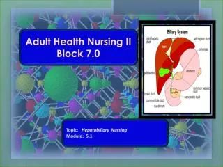
Tracheostomy • Tracheotomy is the surgical incision into the trachea for the purpose of establishing an airway. • Tracheostomy is the stoma, or opening, that results from the procedure of a tracheotomy. • Tracheostomy may be temporary or permanent Block 7.0 Module 4.1
Incision for Trach (Tracheotomy) Block 7.0 Module 4.1
Postoperative Care • #1 priority post-op is airway maintenance & ventilation. • Monitor airway patency, vital signs, hemodynamic status (increased BP, decreased AHR), comfort level. • Assess for complications: • Respiratory distress & hypoxia AEB confusion, restlessness, irritation, agitation, tachypnea, use of accessory muscles & decreased SaO2 (pulse ox) • Hemorrhage: apply direct pressure & summon help • Infection: increased temp & pulse, purulent drainage w/odor, increased redness & tenderness • Wound breakdown common d/t poor nutrition, smoking history, ETOH abuse, wound contamination & previous radiation therapy. Block 7.0 Module 4.1
Carotid Artery Rupture • Extensive surgical wounds in neck area can put carotid artery at risk for rupture. • If leak is suspected, call Rapid Response Team • DO NOT apply pressure could cause immediate rupture • If rupture occurs, apply constant, direct pressure over site & secure airway • Transport patient to OR for resection • Do not leave patient. • Patient at high risk for stroke & death. • To prevent, keep wound dressing wet Block 7.0 Module 4.1
Other Possible Complications Assess for: • Pneumothorax – air in pleural space • Subcutaneous emphysema – crepitus air leak into neck, chest & face tissues if skin is puffy w/crackling sensation, call physician immediately • Bleeding • Infection Block 7.0 Module 4.1
Subcutaneous Emphysema Block 7.0 Module 4.1
Maintaining a Patent Airway • Semi-Fowler’s or high Fowler’s position • Tracheostomy tube (usually temporary) if partial laryngectomy done. Stoma NOT sutured open. • Laryngectomy tube (patient’s only airway) iftotal laryngectomy done. Stoma IS sutured open. Care same as trach tube. Removed 3-6 wks post-op when stoma (surgical opening into trachea) is healed. • Turn, cough and deep breath • Increased mucus secretions -- suction • Humidification (nebulizer) to decrease cough, mucus production, crusting at site • Stoma care: combined wound & airway care Block 7.0 Module 4.1
Maintaining a Patent Airway (cont’d) • Possible complications for tracheostomy tubes: • Tube obstruction from secretions or tube displacement • Tracheostomy tube dislodgment: accidental decannulation. Tube dislodgment in 1st 72h post placement is emergency ventilate patient w/face mask & ambu bag. Call for help. Always have duplicate trach tube, obturator & trach insertion tray at bedside at all times. If >72 hr post-op, use obturator to open site & place new trach tube. Block 7.0 Module 4.1
Temporary Tracheostomy – Tracheostomy Tube • Opening is not sutured open • A tracheostomy tube must always be in place to prevent closure of the opening • Placed for partial laryngectomy & mechanical ventilation temporary airway only pt can still breath through mouth & nose • Has inner & outer cannula inner cannula may be disposable or reusable • Outer cannula may be cuffed or not • Outer cannula may be fenestrated allows pt to speak when capped & inner cannula removed Block 7.0 Module 4.1
Trach Tube, Inner Cannula, Obturator Block 7.0 Module 4.1
Permanent Tracheostomy – Laryngectomy Tube • Placed after total laryngectomy pt’s only airway for life trachea no longer part of oral airway • Opening is sutured open laryngectomy tube can be taken in & out immediately for cleaning or replacement • Prevents shrinkage of stoma until it heals in 3-6 weeks • After open stoma heals, opening is permanent & laryngectomy tube not needed • Not cuffed & has outer cannula only Block 7.0 Module 4.1
Total laryngectomy requires permanent tracheal stoma & laryngectomy tube to maintain airway stoma is sutured open • Results in permanent loss of the voice • Stoma opening is pt’s ONLY airway • No risk for aspiration of food & fluids into lungs since esophagus & trachea are separated • No voice, but normal swallowing Block 7.0 Module 4.1
Laryngectomy Tube & Permanent Stoma Block 7.0 Module 4.1
Trach Suctioning and Care • Suctioning maintains a patent airway and promotes gas exchange. • Assess need for suctioning from the client who cannot cough adequately. -----Trach suctioning (hospital) is strict sterile technique • Always secure tracheostomy tube in place to prevent accidental decannulation • See Craven’s Fundamentals of Nursing, pp. 866-873 Block 7.0 Module 4.1
Complications of Trach Suctioning • Suctioning can cause: • Hypoxia (see causes to follow) • Tissue (mucosal) trauma (see slide) • Infection strict sterile technique never use oral suction equipment to suction an artificial airway • Vagal stimulation results in severe bradycardia & dysrhythmias stop suctioning immediately & oxygenate pt • Cardiac dysrhythmias from hypoxia caused by suctioning stop suctioning & oxygenate pt • See Chart 30-3, p. 584, for Best Practice Block 7.0 Module 4.1
Causes of Hypoxia with Trach Suctioning • Ineffective oxygenation before, during, and after suctioning oxygenate before, during, & after w/100% O2 • Use of a catheter that is too large for the artificial airway standard size is 12 or 14 Fr • Prolonged suctioning time never longer than 10-15 sec. • Excessive suction pressure 80-120 mm/Hg • Too frequent suctioning limit 3 passes Block 7.0 Module 4.1
Prevention of Tissue Damage • Do not apply suction during insertion. • Cuff pressure can cause mucosal ischemia use minimal leak technique. • Check cuff pressure often (<25cm H2O) • Prevent tube friction and movement secure to keep tube mid-line Block 7.0 Module 4.1
Air Warming and Humidification • The tracheostomy tube bypasses the nose and mouth, which normally humidify, warm, and filter the air. • Air must be humidified use humidifier bottle at wall O2 setup Block 7.0 Module 4.1