Download
1 / 64
800 likes | 1.78k Vues
Adult Health Nursing II Block 7.0. …a concept map…. Nursing care planning. Chest Tubes. Placed to drain air & blood from pleural space, restore negative intrapleural pressure & re-expansion of lung Nursing responsibilities (Chart 32-13, p. 648)

E N D
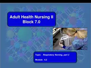
Adult Health Nursing II Block 7.0
…a concept map… Nursing care planning Block 7.0 Module 4.1
Chest Tubes • Placed to drain air & blood from pleural space, restore negative intrapleural pressure & re-expansion of lung • Nursing responsibilities (Chart 32-13, p. 648) • Monitor output hourly call MD if >70ml/hr • Tape tubing junctions • Keep occlusive dressing, sterile 4x4s, and tape at bedside. Use to cover chest puncture site if chest tube falls out. Block 7.0 Module 4.1
Nursing Responsibilities (cont’d) • Assess for tidaling in water seal chamber should be gentle rise & fall of water when pt breaths in & out • Monitor for continuous bubbling in water seal chamber indicates an air leak in the tubing. • Keep 2 padded Kelly forceps at bedside to locate air leak in tubing. • Avoid kinks in the chest tube & drain tubes • Use only sterile water – not normal saline See Craven’s pp. 839-841 Block 7.0 Module 4.1
Chest Tube Indications • Pneumothorax: Air in the pleural space caused by trauma, lung disease, invasive pulmonary procedure, forceful coughing, surgical complication, or may occur spontaneously • To drain air, the chest tube is placed in anterior chest at the second or third intercostal space • Hemothorax: Blood in the pleural space caused by blunt/penetrating trauma or a complication of chest surgery • To drain fluid, the chest tube is placed at lung base • Pleural effusion: Excessive fluid in the pleural space caused by pneumonia, left ventricular heart failure, pulmonary embolism, cancer, or complication of surgery Block 7.0 Module 4.1
Chest Tube Indications • Chylothorax: Accumulation of lymphatic fluid in the pleural space caused by chest trauma, tumor, surgery • Empyema: Pus from an infection, such as pneumonia; must always be drained no matter how small amount • Other considerations: Preventively after cardiac/pulmonary surgery to drain blood postoperatively and prevent cardiac tamponade; also used to instill fluids (chemotherapy, sclerosing agent) Block 7.0 Module 4.1
Types of Chest Drainage Units (CDU) • Chest drainage unit (CDU): Traditional chest drainage unit consists of a collection chamber, water seal chamber, suction control chamber; can drain large amounts of fluid or air • Smaller/lighter portable CDU: Mechanical one-way valve instead of water seal chamber; good for patient who needs drainage only (not suction to reexpand lung), such as noncomplicatedpneumothorax Block 7.0 Module 4.1
Heimlich valve: Contains a one-way flutter valve; air drains out when patient exhales; keep collection device upright and vented to prevent air buildup Examples of underwater conventional CDUs Block 7.0 Module 4.1
Chest Tube Insertion • Done in patient’s room, interventional radiology, or the operating room • Local anesthetic; patient may feel pressure as tube is inserted • Aseptic (sterile) procedure • Patient’s breathing will be easier once lung is re-expanded Block 7.0 Module 4.1
Chest tube insertion • Position patient for comfort depending on site to be inserted • Tube will be anchored with a suture • Insertion site will have an occlusive dressing applied • Connections securely taped • Chest X-ray to confirm position and lung re-expansion Block 7.0 Module 4.1
Risks and complications • Bleeding: Usually minor, but may require surgery if extensive • Infection: Likelihood increases the longer the chest tube is in place • Subcutaneous emphysema (crepitus): Characterized by swelling in face, neck, and chest; crackles on palpation • Lung trauma/bronchopleural fistula: Rare, but patient will have signs and symptoms of respiratory distress, bloody chest tube drainage; tube will be left in place until healed Block 7.0 Module 4.1
Monitor vital signs Assess breath sounds bilaterally Assess the insertion site Encourage the patient to cough Make sure connections are taped securely Keep CDU below the level of the patient’s chest Check water seal and suction control chambers frequently Assess drainage for color Measure drainage at least every 8 hours Document assessment Report immediately bright red blood or red free-flowing drainage >70ml/hour Nursing Considerations Block 7.0 Module 4.1
Care of Chest tube and CDU • Tubing: Avoid loops, aggressive manipulation such as “stripping” or “milking” • Patency: To maintain patency, try “gentle” hand-over-hand squeezing of tubing and release • Clamping: Avoid except when replacing CDU, locating air leak, or assessing when tube will be removed Block 7.0 Module 4.1
Removing the Chest Tube Chest tube can be removed when: • There’s little to no drainage • Air leak is gone • Patient is breathing normally without respiratory distress • Fluctuations in water seal chamber stopped • Chest X-ray shows lung reexpansion with no residual air or fluid Block 7.0 Module 4.1
Procedure for Chest Tube Removal • Gather supplies and explain procedure to patient • The clinician will remove the dressing and sutures • During peak exhalation, the clinician will remove the chest tube in one quick movement • Immediately apply a sterile gauze dressing containing petroleum to prevent air from entering pleural space • Monitor patient’s respiratory status • Arrange for chest X-ray to confirm lung reexpansion • Monitor patient’s respiratory status and SpO2 for 1-2 hours after removal Block 7.0 Module 4.1
Medical Management with Chest Tube Block 7.0 Module 4.1
Acute Respiratory Failure • Not a disease, but a consequence of severe respiratory dysfunction • Lungs are unable to oxygenate blood & remove CO2 adequately, even at rest • Patient is always hypoxic • COPD most common cause. Also: other lung diseases, chest injury, inhalation trauma, neuromuscular disorders, & cardiac conditions Block 7.0 Module 4.1
Acute Respiratory Failure • Critical values of blood gases: • SaO2 <90% • PaO2 <50mm/Hg (hypoxemia) • PaCO2 >50mm/Hg (hypercapnia) • pH <7.35 (acidemia) • Respiratory mechanisms leading to ARF: • Ventilatory failure • Oxygenation failure • Ventilation-perfusion mismatching • Shunting Block 7.0 Module 4.1
Ventilatory and Oxygenation Failure • Ventilatory failure: Gas exchange (ventilation) at the alveolar-capillary membrane is inadequate – too little oxygen reaches the blood and carbon dioxide is retained. • Oxygenation failure: Gas exchange is normal but blood flow (perfusion) to lungs is inadequate. • V/Q (ventilation/perfusion) mismatch occurs. Ventilation &/or perfusion are inadequate. Results in poor respiratory movements & hypoventilation. Block 7.0 Module 4.1
V/Q Scan Mismatch • V/Q scan (ventilation/perfusion scan – obstructed pulmonary blood flow causes V/Q mismatch: an area of lung is ventilated, but not perfused). • Pulmonary embolus most common cause Block 7.0 Module 4.1
Early: Dyspnea Restlessness Fatigue Headache Air hunger Tachycardia Increased blood pressure Decreased breath sounds Use of accessory muscles Later: Confusion Lethargy Tachypnea Central cyanosis Diaphoresis Respiratory arrest S/Sx of Acute Respiratory Failure Block 7.0 Module 4.1
Interventions for ARF • O2 w/pt in high-Fowler’s position • Meds: • Bronchodilators (short-acting) (albuterol) • Corticosteroids (methylprednisolone) • Na bicarb (if metabolic acidosis present) • Anti-anxiety meds (Valium, Ativan) • Diuretics • Antibiotics (if underlying infection present) • May need intubation w/mechanical vent Block 7.0 Module 4.1
Nursing Interventions • Assess/monitor: ABG, SaO2 , VS, respiratory status, neuro status • Establish & maintain airway • Suction if needed • Avoid meds that depress respirations • Turn, cough, deep breath • Prepare for mechanical vent if worsens • Provide emotional support to pt/family Block 7.0 Module 4.1
Acute Respiratory Distress Syndrome (ARDS) (Shock Lung) • Sudden & progressive form of acute respiratory failure w/pulmonary edema, refractory hypoxemia & reduced lung compliance • Inflammation makes damaged alveolar capillary membrane more permeable fluids, protein & blood cells leak into lungs • Loss of surfactant w/decreased elasticity & pulmonary compliance (stiff lung). Alveoli collapse & can be destroyed. Block 7.0 Module 4.1
Acute Respiratory Distress Syndrome (ARDS) • Hypoxia persists even when oxygen at 100% refractory hypoxia • Decreased pulmonary compliance (stiff lung) • 50% mortality rate; incidence >150,000/yr • Most deaths d/t nonpulmonary multiple-system organ failure, often with sepsis Block 7.0 Module 4.1
Risk Factors for ARDS 2 most common causes are: • Gram-negative septic shock • Aspiration of gastric contents (pH 2-4) Other causes: Pneumonia Fat embolus Trauma Drug overdose Near drowning Cardiopulmonary bypass Block 7.0 Module 4.1
“Ground glass” CXR of ARDS Block 7.0 Module 4.1
Early: (12-48h) Cough Restlessness Progressive dyspnea Tachypnea Fine scattered crackles Bilat infiltrates on CXR Progression to late s/sx: Labored dyspnea Confusion Diffuse crackles Tachycardia Hypotension Cyanosis, pallor Profound respiratory distress w/ET intubation S/sx of ARDS Block 7.0 Module 4.1
Diagnostics • ABGs w/lower PaO2 • Refractory hypoxemia w/O2 at 100% • Chest x-ray w/ground-glass appearance • No cardiac involvement on EKG – no left-sided heart failure • Insertion of pulmonary artery cath (Swan-Ganz) to monitor pulmonary artery wedge pressure & cardiac output • Electrolytes, hemoglobin, hematocrit Block 7.0 Module 4.1
Drug Therapy for ARDS • #1 intervention: Oxygen • IV antibiotics if organism identified • Corticosteroids to decrease inflammation, edema • Sedatives, antibiotics, diuretics, inotropics, H2 blockers/PPIs • Many investigational drugs – none has been effective in reducing mortality Block 7.0 Module 4.1
Other Interventions for ARDS • Identify & treat underlying condition • Intubation & mechanical vent w/PEEP (goal: PaO2 >60mmHg, SaO2 >90) • IV fluids for hydration & hypovolemia • Nutritional support via G tube or TPN (35-45 kcal/kg per day) Block 7.0 Module 4.1
Nursing Responsibilities • Assess GI status (prone to GI bleed and stress ulcers w/ARDS & mechanical vent) • Monitor nutrition status • Labs, tube feeding and/or TPN protocols • Daily weights • Hydration status (check BUN) • H2 blockers (Zantac) or PPI (Prilosec) • Infection monitoring: vital signs • Culture results Block 7.0 Module 4.1
Nursing Responsibilities • Fluid volume status • Intake & output • Monitor for dehydration d/t insensible losses • Daily weights • Electrolyte panels • Monitor skin integrity (edema) • Monitor for bleeding Block 7.0 Module 4.1
Mechanical Ventilation • http://www.youtube.com/watch?v=LbMxTk45PTs • Mechanical ventilation is delivered by positive pressure, forcing air into the lungs in one of two ways: 1. Invasively via endotracheal (ET) tube or tracheostomy 2. Noninvasively via mask w/CPAP/BiPAP & iron lung Block 7.0 Module 4.1
Types of Ventilators • Types of ventilators: • Negative-pressure ventilators • Positive-pressure ventilators: used in acute care settings • Pressure-cycled ventilators • Time-cycled ventilators • Volume-cycled ventilators • Microprocessor ventilators Block 7.0 Module 4.1
Iron Lung -- Then Iron Lung -- Now Negative Pressure Ventilator Block 7.0 Module 4.1
Managing Mechanical Ventilation • Normal breathing is controlled by neg pressure system – air is drawn into the lungs • Mechanical ventilation is delivered by positive pressure, forcing air into lungs in one of 2 ways: • Invasively via ET tube or tracheostomy • Noninvasively via mask (CPAP, BiPAP or APAP) • Mechanical ventilation may be lifelong in persons with chronic, progressive neuromuscular diseases that reduce effective ventilation (muscular dystrophy, multiple sclerosis, etc.) Block 7.0 Module 4.1
Modes of Ventilation • The ways in which the client receives breath from the ventilator include: • Assist-control ventilation (AC) • Synchronized intermittent mandatory ventilation (SIMV) • Bi-level positive airway pressure (BiPAP) and others Block 7.0 Module 4.1
ET Tube Placement Block 7.0 Module 4.1
Endotracheal Intubation • ET tube passed through mouth or nose into trachea • Usually temporary for life support (<3 wks) • Provides airway for pts who cannot maintain one on their own, for mechanical vent, & for suctioning lungs • Most often used for hypoxemia & progressive alveolar hypoventilation • Mechanical ventilation may be lifelong in persons with chronic, progressive neuromuscular diseases that reduce effective ventilation (muscular dystrophy, multiple sclerosis, etc.) • Parts of ET tube Block 7.0 Module 4.1
Nursing Responsibilities for Endotrachial Intubation • Verify tube placement: X-ray, assess bilat BS, stomach distention, end-tidal CO2 • Stabilize the tube do not secure to lower jaw d/t to possible movement of jaw & dislodgement of ET tube Block 7.0 Module 4.1
ARDS COPD Thoracic/abdominal surgery Neuromuscular disorders Inhalation injury Status asthmaticus Drug overdose Acute resp failure Pulmonary edema Pulmonary embolus Pneumonia Multiple trauma Shock Multisystem failure Coma Why Patient May Need Mechanical Vent Block 7.0 Module 4.1
Ventilator Modes & Settings • Respiratory rate: Number of breaths ventilator delivers/minute (4 to 20 breaths/minute). • Tidal volume (VT): Volume of gas exchanged with each breath; normal breathing = 7 to 10 ml/kg; ventilator set lower to prevent barotrauma/volutrauma. • Oxygen concentration: Also called fraction of inspired oxygen delivered (FIO2) 22-100% O2. Use lowest FIO2 to maintain SaO2 >90% & PaO2 >60 mm/Hg • Inspiratory/expiratory ratio: The duration of each: ratio usually set for 1:2 to 1:1.5. • Flow rate: speed of tidal volume usually 40L/min • Pressure limit: The maximum pressure a ventilator delivers to achieve tidal volume. Block 7.0 Module 4.1
Nursing Responsibilities • Ventilator management: • Monitor FIO2. ABG’s, CXR, lung sounds • Monitor secretions type and amount • Monitor for pneumothorax • Sedate for ventilator compliance • Monitor ET tube placement • Monitor ventilator settings • Monitor alarm settings • Keep emergency equipment at the bedside. • Assess patient for level of consciousness (LOC), vital signs, lung sounds regularly. • Perform suctioning as needed. • Assess patient’s ability to synchronize breathing with the ventilator. Block 7.0 Module 4.1