Download
1 / 32
600 likes | 1.6k Vues
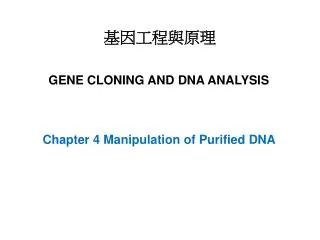
基因工程與原理. GENE CLONING AND DNA ANALYSIS. Chapter 4 Manipulation of Purified DNA. 4 Manipulation of Purified DNA 4.1 The range of DNA manipulative enzymes 4.1.1 Nucleases 4.1.2 Ligases 4.1.3 Polymerases 4.1.4 DNA modifying enzymes 4.2 Enzymes for cutting DNA—restriction endonucleases

E N D
基因工程與原理 GENE CLONING AND DNA ANALYSIS Chapter 4 Manipulation of Purified DNA
4 Manipulation of Purified DNA 4.1 The range of DNA manipulative enzymes 4.1.1 Nucleases 4.1.2 Ligases 4.1.3 Polymerases 4.1.4 DNA modifying enzymes 4.2 Enzymes for cutting DNA—restriction endonucleases 4.2.1 The discovery and function of restriction endonucleases 4.2.2 Type II restriction endonucleases cut DNA at specific nucleotide sequences 4.2.3 Blunt ends and sticky ends 4.2.4 The frequency of recognition sequences in a DNA molecule 4.2.5 Performing a restriction digest in the laboratory 4.2.6 Analyzing the result of restriction endonuclease cleavage Separation of molecules by gel electrophoresis Visualizing DNA molecules in an agarose gel 4.2.7 Estimation of the sizes of DNA molecules 4.2.8 Mapping the positions of different restriction sites in a DNA molecule 4.2.9 Special gel electrophoresis methods for separating larger molecules A few enzymes display multiple activities
4.3 Ligation—joining DNA molecules together 4.3.1 The mode of action of DNA ligase 4.3.2 Sticky ends increase the efficiency of ligation 4.3.3 Putting sticky ends onto a blunt-ended molecule Linkers Adaptors Producing sticky ends by homopolymer tailing 4.3.4 Blunt end ligation with a DNA topoisomerase
4.1.1 Nucleases Figure 4.1 The reactions catalyzed by the two different kinds of nuclease. (a) An exonuclease, which removes nucleotides from the end of a DNA molecule. (b) An endonuclease, which breaks internal phosphodiester bonds.
Figure 4.2 The reactions catalyzed by different types of exonuclease. (a) Bal31, which removes nucleotides from both strands of a double-stranded molecule. (b) Exonuclease III, which removes nucleotides only from the 3′ terminus.
Figure 4.3 The reactions catalyzed by different types of endonuclease. (a) S1 nuclease, which cleaves only single-stranded DNA, including single-stranded nicks in mainly double-stranded molecules. (b) DNase I, which cleaves both single- and double-stranded DNA. (c) A restriction endonuclease, which cleaves double-stranded DNA, but only at a limited number of sites.
4.1.2 Ligases Figure 4.4 The two reactions catalyzed by DNA ligase. (a) Repair of a discontinuity—a missing phosphodiester bond in one strand of a double-stranded molecule. (b) Joining two molecules together.
4.1.3 Polymerases Figure 4.5 The reactions catalyzed by DNA polymerases. (a) The basic reaction: a new DNA strand is synthesized in the 5′ to 3′ direction. (b) DNA polymerase I, which initially fills in nicks but then continues to synthesize a new strand, degrading the existing one as it proceeds. (c) The Klenow fragment, which only fills in nicks. (d) Reverse transcriptase, which uses a template of RNA.
4.1.4 DNA modifying enzymes Figure 4.6 The reactions catalyzed by DNA modifying enzymes. (a) Alkaline phosphatase, which removes 5′-phosphate groups. (b) Polynucleotide kinase, which attaches 5′-phosphate groups. (c) Terminal deoxynucleotidyl transferase, which attaches deoxyribonucleotides to the 3′ termini of polynucleotides in either (i) single-stranded or (ii) double-stranded molecules.
4.2 Enzymes for cutting DNA—restriction endonucleases Figure 4.7 The need for very precise cutting manipulations in a gene cloning experiment. • 1978 Nobel Prizes for W. Arber, H. Smith, and D. Nathans. • The discovery of restriction endonucleases. • One of the key breakthroughs in the development of genetic engineering.
4.2.1 The discovery and function of restriction endonucleases. Figure 4.8 The function of a restriction endonuclease in a bacterial cell: (a) phage DNA is cleaved, but (b) bacterial DNA is not.
4.2.2 Type II restriction endonucleases cut DNA at specific nucleotide sequences 4.2.3 Blunt ends and sticky ends Figure 4.9 The ends produced by cleavage of DNA with different restriction endonucleases. (a) A blunt end produced by AluI. (b) A sticky end produced by EcoRI. (c) The same sticky ends produced by BamHI, BglII and Sau3A.
Figure 4.10 Restriction of the λ DNA molecule. (a) The positions of the recognition sequences for BglII, BamHI, and SalI. (b) The fragments produced by cleavage with each of these restriction endonucleases. The numbers are the fragment sizes in base pairs.
Figure 4.11 Performing a restriction digest in the laboratory.
4.2.6 Analyzing the result of restriction endonuclease cleavage Figure 4.12 (a) Standard electrophoresis does not separate DNA fragments of different sizes, whereas (b) gel electrophoresis does.
Figure 4.13 Visualizing DNA bands in an agarose gel by EtBr staining and ultraviolet (UV) irradiation.
Figure 4.14 Estimation of the sizes of DNA fragments in an agarose gel. (a) A rough estimate of fragment size can be obtained by eye. (b) A more accurate measurement of fragment size is gained by using the mobilities of the HindIII–λ fragments to construct a calibration curve; the sizes of the unknown fragments can then be determined from the distances they have migrated.
Figure 4.15 Using a restriction map to work out which restriction endonucleases should be used to obtain DNA fragments containing individual genes.
Figure 4.16 Restriction mapping. This example shows how the positions of the XbaI, XhoI and KpnI sites on the λ DNA molecule can be determined.
Figure 4.17 The influence of DNA size on migration rate during conventional gel electrophoresis.
Figure 4.18 The difference between conventional gel electrophoresis and orthogonal field alternation gel electrophoresis (OFAGE).
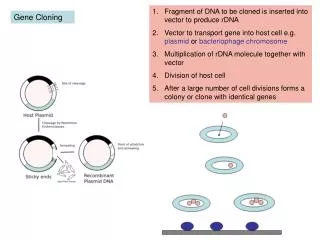
4.3 Ligation—joining DNA molecules together Figure 4.19 Ligation: the final step in construction of a recombinant DNA molecule.
4.3.2 Sticky ends increase the efficiency of ligation Figure 4.20 The different joining reactions catalysed by DNA ligase: (a) ligation of blunt-ended molecules; (b) ligation of sticky-ended molecules.
4.3.3 Putting sticky ends onto a blunt-ended molecule Figure 4.21 Linkers and their use: (a) the structure of a typical linker; (b) the attachment of linkers to a blunt-ended molecule.
Figure 4.22 A possible problem with the use of linkers. Compare this situation with the desired result of BamHI restriction, as shown in Figure 4.21(b).
Figure 4.23 Adaptors and the potential problem with their use. (a) A typical adaptor. (b) Two adaptors could ligate to one another to produce a molecule similar to a linker, so that (c) after ligation of adaptors a blunt-ended molecule is still blunt-ended and the restriction step is still needed.
Figure 4.24 The distinction between the 5′ and 3′ termini of a polynucleotide.
Figure 4.25 The use of adaptors: (a) the actual structure of an adaptor, showing the modified 5′-OH terminus; (b) conversion of blunt ends to sticky ends through the attachment of adaptors.
Figure 4.26 Homopolymer tailing: (a) synthesis of a homopolymer tail; (b) construction of a recombinant DNA molecule from a tailed vector plus tailed insert DNA; (c) repair of the recombinant DNA molecule.
4.3.4 Blunt end ligation with a DNA topoisomerase Figure 4.27 The mode of action of a Type 1 DNA topoisomerase, which removes or adds turns to a double helix by making a transient break in one of the strands.
Figure 4.28 Blunt end ligation with a DNA topoisomerase. (a) Cleavage of the vector with the topoisomerase leaves blunt ends with 5′-OH and 3′-P termini. (b) The molecule to be cloned must therefore be treated with alkaline phosphatase to convert its 5′-P ends into 5′-OH termini. (c) The topoisomerase ligates the 3′-P and 5′-OH ends, creating a double-stranded molecule with two discontinuities, which are repaired by cellular enzymes after introduction into the host bacteria.