Insect Biochemistry - Excretion
Insect Biochemistry - Excretion. Kuang-Hui Lu Department of Entomology National Chung Hsing University. CONTENTS. Introduction The Malpighian tubules Ultrastructure of Malpighian tubule cells Formation of primary urine in Malpighian tubules

Insect Biochemistry - Excretion
E N D
Presentation Transcript
Insect Biochemistry- Excretion Kuang-Hui Lu Department of Entomology National Chung Hsing University
CONTENTS • Introduction • The Malpighian tubules • Ultrastructure of Malpighian tubule cells • Formation of primary urine in Malpighian tubules • A proton pump is the driving mechanism for urine formation • Selective reabsorption in the hindgut • The role of the excretory system in maintaining homeostasis • Cryptonephridial systems
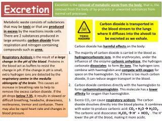
Introduction • Excretion: any process that eliminates the interaction of harmful substances with cells and tissues. • eliminate nitrogenous metabolites • maintain ions and water balance • remove ingested chemicals – e.g. allelochemicals
Introduction • Excretory organs of insects • Malpighian tubules – collect a filtrate from the hemolymph and pass this primary urine to the hindgut. • Hindgut – secrete additional components into the secreta, and reabsorbe some substances into the hemolymph.
Introduction • The major function of excretory system is to maintain the internal environment of an organism by separating and eliminating metabolic wastes and other toxic substances. • Because metabolic wastes are often dissolved in water, excretory processes are also closely associated with osmoregulation and the maintenance of water balance.
Fig. A generalized scheme of excretion showing the collection of fluid in the Malpighian tubules, and extensive reabsorption of water, K+, and useful substances from the hindgut, primarily the rectum.
Malpighian Tubules • Malpighian tubules are primary excretory organs of insects • Malpighian tubules are long, tubular structures, usually arising at the junction of the mid- and hindgut and terminating blindly in the hemocoel. • The tubules vary in number from 2 to more than 100 in various insect species. • Tracheal connections to Malpighian tubules are numerous. • A small spiral muscle frequently runs along the surface of a tubule. (next slide)
Malpighian Tubules • The Malpighian tubules arise during embryogenesis as evaginations of the gut, usually originating at the junction of the midgut and hindgut. • The tubule walls consist of a single cell layer of epithelial cells and are differentiated by structure and function along the length of the tubule.
Malpighian Tubules • The process of excretion is a two-step process, with much of the fluid that is taken up by the tubules resorbed by the hindgut before is passes out of the body.
Ultrastructure of Malpighian Tubule Cells • A single layer of epithelial cells surrounds the lumen of a tubule. • Several different cell types have been identified, but their specific functions have not been elucidated in many cases. • Type 1 (or principal tubule cells) – tubule cells have a brush border of microvilli on the apical surface. (next slide)
Transport of Substances Through the Malpighian Tubule Cells • The primary urine formed in the lumen of the Malpighian tubules is a filtrate of the hemolymph, and it contains most of the small ions and molecules that occur in the hemolymph.
Transport of Substances Through the Malpighian Tubule Cells • The urine:hemolymph concentration ratio for many of the filtered substances approaches unity, indicating passive movement across the tubule cell membranes. • But some components are actively secreted and their urine:hemolymph ratio is always greater than one.
Primary Urine Formation • Urine formation in Malpighian tubules mainly relies on a proton pump in the apical membrane of Malpighian tubule cells that actively secretes protons (H+) into the tubule lumen against an electrochemical gradient. • The pump causes the tubule lumen to become positive to the hemolymph, and creates highly variable gradients in pH across the apical membrane of principal cells. • The proton gradient provides the energy for an antiporter mechanism that exchanges K+ for H+ across the apical membrane. (next slide)
Primary Urine Formation • Secretion of cations (H+, Na+, and K+) across the apical membrane appears to be electrically coupled with Cl- transport in the basolateral membrane of tubule cells. (next slide) • The formation of urine volume is highly dependent on the K+ concentration in hemolymph or saline. (next slide) • The process driven by the proton pump has been called a standing gradient process. • Additional processes might be involved in substances interring the tubule lumen. (next slide)
Hormonal Control of Urine Formation • The rates of urine formation and ion secretion are controlled by diuretic hormones and certain non-peptide compounds, such as 5-hydroxytryptamine (5-HT or serotonin) • The diuretic neuropeptides isolated from insects fall into one of two hormone families: • Corticotropin-releasing factor (CRF)-related peptides: range in size from 30-46 amino acids; has approximately 30% sequence homology with the CRF family of vertebrate peptides. • Insect kinins: small peptides of between 6 and 15 amino acids
Urine Formation • The primary urine formed by the Malpighian tubules is isosmotic or sometimes slightly hyposmotic to the hemolymph. • Malpighian tubules are not capable of producing primary urine that is appreciably hyperosmotic to the hemolymph. • The hindgut proceeds to concentrate waste products by reabsorbing water and useful substances.
The Bioassay of Malpighian Tubule Function Devised by Ramsay By analyzing the primary urine formed in the droplets, it was discovered that it was isosmotic with the hemolymph, but with potassium concentrations up to 20 times higher.
Arrangement for Experimental Perfusion of an Isolated Tubule
The Cumulative Formation of Primary Urine by an Isolated Tubule
Anatomical Specialization of Hindgut Epithelial Cells • The hindgut is the second system that completes the excretion process by • selectively reabsorbing some substances into the hemolymph • leaving others in the lumen • actively secreting some substance into the hindgut lumen • The rectal cuticular lining has greater permeability than the cuticular lining on foregut cells. • The epithelial cells of the hindgut are specialized for both active secretion and active reabsorption.
Anatomical Specialization of Hindgut Epithelial Cells • Rectal cells (or rectal pad cells, rectal papillae cells) – a group cells in the rectum that have special modifications for reabsorption. (next slide) • In Diptera, the cells of a rectal papilla are large, usually cuboidal cells that surround a central channel in the papilla that opens into the hemolymph space through a valve. (next slide).
A Rectal Cell and Its Ion Transport • The rectum consists of the enlarged posterior-most section of the hindgut, often containing specialized structures called papillae or rectal pads that are enlarged epithelial cells. • The rectum transports water and ions from the material within the gut lumen into the hemolymph.
Secretion and Reabsorption in the Ileum • The ileum is the most anterior part of the hindgut, occurring just posterior to the origin of the Malpighian tubules in most insects. • In locust S. gregaria, the ileum is a major site for • isosmotic fluid reabsorption • active Na+ and Cl- reabsorption • active secretion of proline as an energy source
Secretion and Reabsorption in the Ileum • The driving mechanism for ion and water reabsorption in the ileum is an electrogenic Cl- pump. • A neuropeptide, the ion transport peptide (ITP) stimulates Na+, Cl- and water reabsorption, and promotes passive reabsorption of K+ by electrical coupling. • The ileum plays a major role in acid-base balance by secretion of H+ into the lumen, formation of NH4+, and reabsorption of HCO3-.
Reabsorption in the Rectum • The rectum is the final and major site for reabsorption of ions, water, and nutrients. • It is capable of reabsorbing fluid against strong osmotic gradients, ultimately producing a very concentrated hyperosmotic excreta in many insects. • The driving mechanism for cation and water reabsorption, as in the ileum, is an electrogenic Cl- pump under the influence of a neuropeptide hormone, chloride transport stimulating hormone (CTSH), from the corpora cardiaca.
Fig. Ions are transported in and out of locust rectal cell by numerous mechanisms.
Electrolyte Homeostasis • In mosquito A. aegypti, feeding on a blood meal stimulates the release of mosquito natriuretic peptide (MNP) from the CC, and cAMP is produced and acts selectively to open Na+ channels in the basolateral membrane of the Malpighian tubule cells. • Movement of water into tubule cells follows the osmotic gradient. • The ion flex generated by MNP and cAMP is specifically an increase in secretion of Na+. K+ movement is not influenced. • The Cl- load from the blood meal move from hemolymph to tubule lumen in a passive transport pathway between the cells (paracellular pathway).
Electrolyte Homeostasis • Larval A. aegypti live in fresh water, and in response to an increase in salinity • Secrete 5-hydroxytryptamine (serotonin) into the hemolymph • Increase cAMP in the Malpighian tubules • Serotonin and cAMP stimulate fluid and ion (Na+ and K+) secretion rates in the tubules, but urine is not concentrated with respect to the ions
Electrolyte Homeostasis • Beyenbach (1995) has reviewed three potential physiological processes through which A. aegypti may regulate rates of ion and fluid excretion • The proton pump that supplies energy for Na+ and K+ secretion to the tubule lumen • The resistance Rc across the tubule cells that control ion channels in the basolateral membrane • The resistance of the passive transport pathway for Cl- movement
Water Homeostasis • Water excretion and retention are regulated by hormones. • Diuretic hormones promote fluid formation and rapid excretion by the Malpighian tubules • Corticotropin-releasing factor (CRF)-related peptides: range in size from 30-46 amino acids. • Insectkinins: small peptides of between 6 and 15 amino acids. • Antidiuretic hormones act on the hindgut and promote water reabsorption • Chloride transport-stimulating hormone (CTSH) • Ion transport peptide (ITP)
A Filter Chamber • In some Homopterans that feed exclusively on plant juices containing low concentrations of nutrients, the digestive tract forms an arrangement known as a filter chamber.
Acid-Base Homeostasis • The excretory system is important in maintaining the acid-base balance of body fluids and tissues. • Acid-base regulation in S. gregaria • Secretion of H+ and formation of NH4+ in the ileum is a principal mechanism for excreting excess acid equivalents. • The ileum is a major site of ammoniagenesis in locusts in which hindgut cells specifically metabolize amino acids and glucose for energy.
Acid-Base Homeostasis • Excretion of total ammonia nitrogen serves several functions in locusts • Ammonium urate (i.e. NH3 reacts with uric acid) allows the insect to conserve Na+ • Conversion of NH3 to NH4+ in the ileal cells is equivalent to removal of H+ • Excretion of NH3 by locusts conserves water • Increases nitrogen excretion by 25% more than excretion of only Na- or K-urate.
Nitrogen Homeostasis • The metabolism of proteins and nucleic acids produce ammonia. • Some of this ammonia can be recycled into amino acid synthesis by the formation of glutamate from a-ketoglutarate and glutamine from glutamate. • The excess ammonia that remains is highly toxic unless it is diluted with water. • High levels of ammonia can interrupt nervous transmission by substituting for necessary potassium and can also alter carbohydrate and lipid metabolism.
The Incorporation of Ammonia for the Synthesis of Amino Acids
Nitrogen Homeostasis • Organisms must have excretory systems to avoid the toxic accumulation of ammonia. • Because ammonia is very soluble in water, its concentrations have to be maintained below levels that are toxic. • Most terrestrial organisms have taken the pathway of the incorporation of the nitrogen into either urea or uric acid, which can be concentrated in body fluid to a much greater extent than can ammonia and require less water for dilution.
Nitrogen Homeostasis • In insects, the need for water conservation may have been the driving force for the incorporation of their nitrogen wastes into uric acid. • The fat body is the primary site for uric acid synthesis. • Uric acid does not dissolve well in water and therefore fails to reach toxic levels in body fluids, so it requires about 50 times less water to dilute than does ammonia. • Insolubility of uric acid in water allows it to be excreted in a dry form without having a significant effect on water balance.
Nitrogen Homeostasis • Insects pay a high price for the benefits they derive from employing uric acid as a way to excrete nitrogen and still maintain a positive water balance. • The synthesis of uric acid results loss of several carbon atoms. • Eight ATP are required to first make the intermediary metabolite, inosine monophosphate (IMP)
Xanthine dehygrogenase Fig. Pathway for the synthesis of uric acid from nucleic acids and protein.
Release of Ammonia by the Deamination of Amino Acids e.g. blowfly larvae and some cockroaches and locusts.
Storage Excretion • Because uric acid is so insoluble, it can be easily stored without it interacting with other physiological processes. • Some cockroaches accumulate up to 10% of their dry weight in uric acid stored in specialized urate cells in the fat body, which can be utilized during periods of dietary stress. • In some lepidoptera, the fat body shifts from excretion of uric acid to its storage during the last larval instar.
A Cryptonephridial Complex • Many families of Coleoptera, Lepidoptera and some saw-fly larvae, that live under extremely dry conditions, the ends of the Malpighian tubules do not lie free in the hemocoel. • Instead, the terminal segments of the tubules are closely associated with the wall of the rectum in what is called a cryptonephridial complex. (next slide) • It appears to be an arrangement that enables very efficient conservation of water.
A Cryptonephridial Complex • The cryptonephridial complex is found in most lepidopteran larvae and many coleopterans. • The cryptonephridial complex performs two functions: • Resorb water from the hindgut very efficiently. (next slide) • In some insects is able to absorb atmospheric water from the humidity in the hindgut.
Fig. An SEM photo of the small muscle (arrow) that often spirals along the length of a Malpighian tubule of some insects.
A Cross Section through the Primary Type of Malpighian Tubule
Fig. The general structure of a Malpighian tubule cell from the proximal tubule segment of the last instar of Drosophila melanogaster that illustrates extensive basal infoldings, a relatively short path across the narrow cell, and long microvilli on the apical surface of the cells.