Download
1 / 81
820 likes | 991 Vues
Discover the diverse world of protozoa, from their unicellular nature to their medical importance as parasites. Learn about the different types, feeding habits, and medically relevant species. Explore key topics like amoebas, flagellates, and apicomplexans.

E N D
Chapter 12: Characterizing and Classifying Eukaryotes Protozoa, Fungi, Algae, and Parasites
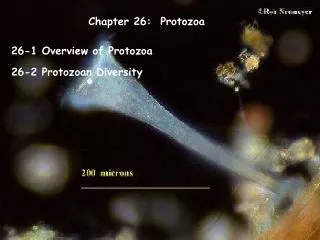
CLASSIFICATION OF PROTOZOA • Unicellular, chemoheterotrophic, eucaryotic organisms of kingdom Protista (3-2000 mm). • Protozoan means “first animal”. • 20,000 species, only a few are pathogens. • Most are free-living organisms that inhabit water and soil. Some live in association with other organisms as parasites or symbionts. • Reproduce asexually by fission, budding, or schizogony. • Some exhibit sexual reproduction (e.g.: Paramecium). • Trophozoite: Vegetative stage which feeds upon bacteria and particulate nutrients. • Cyst: Some protozoa produce a protective capsule under adverse conditions (toxins, scarce water, food, or oxygen).
PROTOZOA (Continued) Nutrition • Most are heterotrophic aerobes. Intestinal protozoa can grow anaerobically. • Some ingest whole algae, yeast, bacteria, or smaller protozoans. Others live on dead and decaying matter. Parasitic protozoa break down and absorb nutrients from their hosts. • Some transport food across the membrane. • Others have a protective covering (pellicle) and required specialized structures to take in food. • Ciliates take in food through a cytostome. • Digestion takes place in vacuoles. • Waste may be eliminated through plasma membrane or an anal pore.
Medically Important Protozoa 1. Amoebas (Phylum Sarcodina) • Move and feed by extending projections (pseudopods) • Engulf food with pseudopods and carry out phagocytosis • Several species cause amoebic dysenteries of varying degrees of severity. • Entamoebahystolytica: Feeds on red blood cells. Produces dysentery and extraintestinal cysts. • Dientamoebafragilis: Found in 4% of humans. Usually commensal. Can cause chronic, mild diarrhea. • Other diseases include: • Meningoencephalitis: Caused by Naegleriafowleri. Penetrate nasal mucosa of swimmers in warm waters. Mortality rate almost 100%. • Keratitis: Caused by Acanthamoeba. Can cause blindness. Associated with use of contact lenses.
2. Flagellates (Phylum Mastigophora) • Move by one or more whiplike flagella. Some parasitic flagellates have up to eight flagella. • Most are spindle shaped with flagella projecting from anterior end. • Outer membrane is a tough pellicle. Food is ingested through an oral groove or cytosotome. • Important pathogens: • Trichomonasvaginalis: Causes genital and urinary infections. Has undulating membrane. Lacks a cyst stage. Transmitted sexually or by fomites. • Giardialamblia: Causes a persistent intestinal infection (giardiasis) with diarrhea, nausea, flatulence, and cramps. In U.S. most common cause of waterborne diarrhea. About 7% of U.S. population are healthy carriers. • Trypanosomabruceigambiense: Hemoflagellate (blood parasite). Causes African sleeping sickness. Transmitted by Tse-Tse fly. • Trypanosomacruzi: Hemoflagellate that causes Chaga’s disease, a cardiovascular disease common in Texas and Latin America. Transmitted by kissing bug living in mud houses or blood transfusions. Can cause heart damage (heart failure, arrythmia over several years).
Medically Important Protozoa (Continued) 3. Apicomplexans (Phylum Sporozoa) • Not motile in their mature form. • Obligate intracellular parasites. • Have specialized organelles at tip (apex) of cells that allow them to penetrate host tissues. • Complex life cycles. May have more than one host. Definitive host: Harbors sexually reproducing form. Intermediate host: In which asexual reproduction occurs.
Medically Important Protozoa (Continued) 3. Apicomplexans • Important pathogens: • Plasmodium vivaxand falciparum: Cause malaria in humans (intermediate host). Initially treated with quinine, drug resistance is a major problem today. Major cause of worldwide mortality: Kill 3 million people/year and infect 500 million. Transmitted by Anophelesmosquito (definitive host). DDT was used extensively in 1960s in an attempt to eradicate the mosquito vector. Successful vaccine not available yet.
Life Cycle of Plasmodium spp. the Infectious Agent of Malaria
Medically Important Protozoa (Continued) 3. Apicomplexans • Important pathogens: • Toxoplasmagondii: Causes toxoplasmosis in humans. Causes blindness and lymphatic infections in adults. Dangerous to pregnant women, causes severe neurological defects in unborn children. Cats are part of life cycle, oocysts excreted in feces. Contact with infected feces or meat are means of transmission. • Cryptosporidium: Causes respiratory and gallbladder infections in immunosuppressed individuals. Found in intestines of mammals and water. Major cause of death in AIDS patients. • Cyclosporacayetensis: New parasite (1996) caused diarrhea associated with raspberries.
Medically Important Protozoa (Continued) 4. Ciliates (Phylum Ciliophora) • Move and obtain food using cilia. • Only known human pathogen is Balantidium coli, which causes a severe intestinal infection in pigs and humans.
Paramecium caudatum is a Ciliated ProtozoanConjugation Between Opposite Mating Strains
CLASSIFICATION OF FUNGI (Mycology) • Diverse group of heterotrophs • Many are ecologically important saprobes or saproprophytes (consume dead and decaying matter) • Others are parasites. • Most are multicellular, but yeasts are unicellular • Most are aerobes or facultative anaerobes • Cell walls are made up of chitin (polysaccharide). • Over 100,000 fungal species identified. Only about 200 are human or animal pathogens • Most human fungal infections are nosocomial and/or occur in immunocompromised individuals (opportunistic infections). • Fungal diseases in plants cause billions of dollars a year in losses
CHARACTERISTICSOFFUNGI 1. Yeasts • Unicellular fungi, nonfilamentous, typically oval or spherical cells. Reproduce by mitosis: • Fission yeasts: Divide evenly to produce two new cells (Schizosaccharomyces). • Budding yeasts: Divide unevenly by budding (Saccharomyces). Budding yeasts can form pseudohypha, a short chain of undetached cells. Candida albicans invade tissues through pseudohyphae. • Yeasts are facultative anaerobes, which allows them to grow in a variety of environments. • When oxygen is available, they carry out aerobic respiration. • When oxygen is not available, they ferment carbohydrates to produce ethanol and carbon dioxide.
CHARACTERISTICSOFFUNGI (Continued) 2. Molds and Fleshy Fungi • Multicellular, filamentous fungi. • Identified by physical appearance, colony characteristics, and reproductive spores. • Thallus: Body of a mold or fleshy fungus. Consists of many hyphae. • Hyphae (Sing: Hypha): Long filaments of cells joined together. • Septate hyphae: Cells are divided by cross-walls (septa). • Coenocytic (Aseptate) hyphae: Long, continuouscells that are not divided by septa. Hyphae grow by elongating at the tips. Each part of a hypha is capable of growth. • Vegetative Hypha: Portion that obtains nutrients. • Reproductive or Aerial Hypha: Portion connected with reproduction. • Mycelium: Large, visible, filamentous mass made up of many hyphae.
CHARACTERISTICSOFFUNGI (Continued) Dimorphic Fungi • Can exist as both multicellular fungi (molds) and yeasts. • Many pathogenic species. • Mold form produces aerial and vegetative hyphae. • Yeast form reproduces by budding. • Dimorphism in pathogenic fungi typically depends on temperature: • At 37oC: Yeast form. • At 25oC: Mold form. • Dimorphism in nonpathogenic fungi may depend on other factors: Carbon dioxide concentration.
LIFECYCLEOFFUNGI • Filamentous fungi can reproduce asexually by fragmentation of their hyphae. • Fungal spores are formed from aerial hyphae and are used for both sexual and asexual reproduction. 1. Asexual spores: Formed by the aerial hyphae of one organism. New organisms are identical to parent. • Conidiospore: Unicellular or multicellular spore that is not enclosed in a sac. • Chlamydospore: Thick-walled spore formed within a hyphal segment. • Sporangiospore: Asexual spore formed within a sac (sporangium). 2. Sexual spores: Formed by the fusion of nuclei from two opposite mating strains of the same species. New organisms are different from both parents.
IMPORTANTDIVISIONSOFFUNGI 1. Division Zygomycota (Conjugation Fungi) • Over 1100 species known, most are saprophytes • Also known as bread molds • Molds with coenocytichyphae (lack septa) • Asexual Reproduction: Used most of the time Sporangiospore: Asexual spore enclosed within a sporangium or balloon-like sac at the end on an aerial hypha • Sexual Reproduction: Occurs through conjugation, the joining of hypha of two different strains (plus and minus) Zygospores: Sexual spores are enclosed in a thick wall. • Generally not pathogens • Rhizopusnigricans: Common black bread mold. May cause opportunistic infections in diabetes patients
Life Cycle of a Zygomycete: Black Bread Mold (Rhizopus)Reproduces Asexually and Sexually
Reproductive Structures of Zygomycete (Rhizopus)Sporangia (asexual) and Zygospore (sexual)
2. Division Ascomycota (Sac Fungi) • Molds with septatehyphae and some yeasts. • Asexual Reproduction: Conidia means dust. Conidiosporesnot enclosed in a sac. Become airborne easily. Form chains (broom-like structures). • Sexual Reproduction: Ascospores enclosed in a sac-like structure (ascus). • Include common antibiotic producing fungi and yeasts, and several human pathogens. • Penicilliumnotatum(Produces penicillin) • Saccharomyces(Brewer’s yeast) • Trychophyton(Athlete’s foot) • Aspergillus(Carcinogenic aflatoxin in peanuts), • Blastomyces(Respiratory infections) • Histoplasmacapsulatum(Respiratory and systemic infections)
Life Cycle of Eupenicillium (Ascomycete)Reproduces Asexually and Sexually
Athlete’s Foot (Tinea pedis) Source: Doctorfungus Corporation, 2000
Severe nail infection with Trichophyton rubrum in a 37-year-old male AIDS patient. Source: Intern. J. Dermatol. 31(1992): 453.
Disseminated Histoplasma capsulatum, skin infection. Source: Microbiology Perspectives, 1999.
3. Division Basidiomycota (Club Fungi) • Have septatehyphae. • Include mushrooms, toadstools, rusts, and smuts. • Sexual Reproduction: Produce basidiospores: Spores formed externally on a club shaped sexual structure or base called basidium. • Asexual Reproduction: Through hyphae. • Examples: • Cryptococcus neoformans: Causes opportunistic respiratory and CNS infections in AIDS patients. • Amanita: Mushroom produces lethal toxins to humans. • Clavicepspurpurea: Produces ergot toxin in wheat and rye.
Life Cycle of a BasidiomyceteMushrooms are Produced Sexually
IMPORTANTDIVISIONSOFFUNGI 4. Division Deuteromycota • Most are closely related to Ascomycota, division abandoned by most taxonomists • Not known to produce sexual spores • Reproduce asexually • Candida albicans: Causes yeast infections of vagina in women. Opportunistic infections of mucous membranes in AIDS patients. • Pneumocystiscarinii: Causes pneumonia in AIDS patients. Leading cause of death in AIDS patients. Originally classified as a protozoan.
Opportunistic Infection by Candida albicans in an AIDS Patient Source: Atlas of Clinical Oral Pathology, 1999
Other Fungi Microsporans • Reclassified as fungi in 2003. • Obligate intracellular parasites, lack mitochondria and microtubules. • Discovered in 1984 to cause chronic diarrhea and conjunctivitis, mainly in immunocompromised (AIDS, cancer, etc.) patients.
NUTRITIONALADAPTATIONSOFFUNGI Fungi absorb their food, rather than ingesting it. • Fungi grow better at a pH of 5, which is too acidic for most bacteria. • Almost all molds are aerobic. Most yeasts are facultative anaerobes. • Fungi are more resistant to high osmotic pressure than bacteria. • Fungi can grow on substances with very low moisture. • Fungi require less nitrogen than bacteria to grow. • Fungi can break down complex carbohydrates (wood, paper), that most bacteria cannot.
FUNGALDISEASES Mycosis:Any fungal disease. Tend to be chronic because fungi grow slowly. Mycoses are classified into the following categories: I. Systemicmycoses: Fungal infections deep within the body. Can affect a number if tissues and organs. • Usually caused by fungi that live in the soil and are inhaled. Not contagious. • Examples: • Histoplasmosis (Histoplasma capsulatum): Initial infection in lungs. Later spreads through blood to most organs. • Coccidiomycosis (Coccidioides immites): Resembles tuberculosis.
Systemic Mycosis: Histoplasmosis Disseminated Histoplasma capsulatum, lung infection. Source: Microbiology Perspectives, 1999.
FUNGALDISEASES (Continued) II. Cutaneousmycoses: Fungal infections of the skin, hair, and nails. • Secrete keratinase, an enzyme that degrades keratin. • Infection is transmitted by direct contact or contact with infected hair (hair salon) or cells (nail files, shower floors). • Examples: • Ringworm (Tinea capitis and T. corporis) • Athlete’s foot (Tinea pedis) • Jock itch (Tinea cruris)
Cutaneous Mycosis Ringworm skin infection: Tinea corporis Source: Microbiology Perspectives, 1999
Cutaneous Mycosis: Jock Itch Organsim: Tinea cruris Source: DermNet.com
Cutaneous Mycosis Candida albicans infection of the nails. Source: Microbiology Perspectives, 1999.
FUNGALDISEASES (Continued) III. Subcutaneousmycoses: Fungal infections beneath the skin. • Caused by saprophytic fungi that live in soil or on vegetation. • Infection occurs by implantation of spores or mycelial fragments into a skin wound. • Can spread to lymph vessels. IV. Superficial mycoses: Infections of hair shafts and superficial epidermal cells. Prevalent in tropical climates.
FUNGALDISEASES (Continued) Opportunistic mycoses: Caused by organisms that are generally harmless unless individual has weakened defenses: • AIDS, cancer, transplant, and diabetic patients • Individuals treated with broad spectrum antibiotics • Very old or very young individuals (newborns). • Examples: • Aspergillosis: Inhalation of Aspergillus spores. • Yeast Infections or Candidiasis: Caused mainly by Candida albicans. Part of normal mouth, esophagus, and vaginal flora.
ECONOMIC IMPORTANCE OF FUNGI • 25-50% of harvestedfruits and vegetables are damaged by fungi. • Fungal infections of plants are commonly called rots, rusts, blights, wilts, and smuts. • Beneficial fungi: • Candida oleophila: Prevents fungal growth on harvested fruits. • Saccharomyces cerevisiae: Used to make bread and wine. • Genetically engineered yeast strains are used to make proteins (Hepatitis B vaccine). • Taxomyces: Produces anticancer drug taxol. • Trichoderma: Produces cellulase. Used to make fruit juice. • Saccharomyces boulardi: Used as probiotic because it kills other yeast.
II. ALGAE • Most are simple eucaryotic photosynthetic autotrophs. • Unicellular or multicellular. Kingdom Protista. • Most are found in the ocean or other bodies of water. Need water for support, reproduction, and nutrition. • Absorb nutrients from the water over entire surface. • Reproduction: All reproduce asexually. Some can also reproduce sexually. • Most are not pathogens. A few produce toxins that are harmful to humans.
II. ALGAE • Vegetative Structures of multicellular algae: • Thallus: Body. Lacks conductive tissue. • Holdfasts: Anchor alga to rock. • Stipes: Hollow, stem-like structures. Does not support weight. • Blades: Leaf-like structures. • Pneumatocyst: Floating, gas-filled bladder.
DIVISIONS OF ALGAE • Green algae (Chlorophyta): May be unicellular or multicellular. Have cellulose cell walls, contain chlorophyll a and b, and store sugar and starch like plants. Most are microscopic. Live close to water surface. Believed to be the ancestors of terrestrial plants. • Brown Algae or Kelp (Phaeophyta): Macroscopic (up to 50 m long). Most are found in coastal waters, at intermediate depths. Rapid growth. Can be harvested regularly.
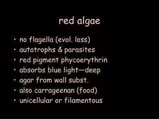
DIVISIONS OF ALGAE • Red Algae (Rhodophyta): Live at greater ocean depths than other algae. Red pigments allow them to absorb blue light that penetrates deepest into ocean. Agar is extracted from many red algae. Some produce lethal toxins. • Source of gel-like polysaccharides agar and carragean.
Golden, Yellow-Green Algae, and Diatoms (Chrysophyta) • Diatoms: Unicellular or filamentous algae with complex cell walls with silica or calcium. • Twoparts of cell wall fit together like Petri dish. Distinctive patterns are used for identification. Store energy in form of oil. • Some diatoms can cause neurological disease (memory loss and diarrhea) in people who eat mussels, due to domoic acid intoxication. • Fossil deposits of diatoms (diatomaceous earth) are used as filtering agents and abrasives in several industries.
Dinoflagellates (Plankton): Unicellular free-floating algae. Rigid structure due to cellulose in plasma membrane. Some dinoflagellates produce neurotoxins, which kill fish, marine mammals, and humans. • Paralytic shellfish poisoning: Consumption of clams and mussels that have eaten dinoflagellates (Alexandrium spp.) that produce neurotoxin. • Red Tide: Caused by large concentrations of Alexandrium. Avoid harvesting mollusks and fish during red tide. • Euglenoids: Unicellular, flagellated algae. Semi-rigid plasma membrane (pellicle). Most have anterior red eye spot. Frequently studied with protozoa, because lack a cell wall.