Download
1 / 24
330 likes | 765 Vues
ANATOMY OF THE SHOULDER REGION. Dr. Ahmed Fathalla Ibrahim & Dr. Zeenat Zaidi. OBJECTIVES. At the end of the lecture, students should: List the name of muscles of the shoulder region.

E N D
ANATOMY OF THE SHOULDER REGION Dr. Ahmed Fathalla Ibrahim & Dr. ZeenatZaidi
OBJECTIVES At the end of the lecture, students should: • List the name of muscles of the shoulder region. • Describe the anatomy of muscles of shoulder region regarding: attachments of each of them to scapula & humerus, nerve supply and actions on shoulder joint • List the muscles forming the rotator cuff and describe the relation of each of them to the shoulder joint. • Describe the anatomy of shoulder joint regarding: type, articular surfaces, stability, relations & movements.
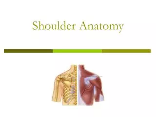
MUSCLES OF SHOULDER REGION Posterior view • These muscles connect scapula to humerus (move humerus through shoulder joint). • Deltoid. • Supraspinatus. • Infraspinatus. • Teres minor. • Teres major. • Subscapularis. 2 1 3 4 5 Anterior view 6
DELTOID • A triangular muscle, forms the contour of the shoulder. • Origin: lateral 1/3 of clavicle + acromion and spine of scapula (look to insertion of trapezius). • Insertion: deltoid tuberosity of humerus. • Nerve supply: axillary nerve. • Actions: • Anterior fibers: flexion & medial rotation of humerus (arm, shoulder joint). • Middle fibers: abduction of humerus from 15° - 90 °. • Posterior fibers: extension & lateral rotation of humerus.
SUPRASPINATUS & INFRASPINATUS • Origin: • Supraspinatus: supraspinousfossa. • Infraspinatus: infraspinaousfossa. • Insertion: greater tuberosity of humerus. • Nerve supply: suprascapular nerve. • Action: • Supraspinatus: abduction of humerus from 0° - 15°. • Infraspinatus: lateral rotation of humerus. Supraspinatus Infraspinatus
TERES MINOR • Origin: lateral border of scapula • Insertion: greater tuberosity of humerus. • Nerve supply: axillary nerve. • Action: lateral rotation of humerus.
TERES MAJOR • Origin: lateral border of scapula • Insertion: bicipital groove of humerus (look to insertion of latissimusdorsi & pectoralis major). • Nerve supply: lower subscapular nerve. • Actions: extension, adduction & medial rotation of humerus (look to action of latissimusdorsi).
SUBSCAPULARIS • Origin: subscapularfossa. • Insertion: lesser tuberosity of humerus. • Nerve supply: upper & lower subscapular nerves. • Action: medial rotation of humerus.
SHOULDER (Glenohumeral) JOINT TYPE: Synovial, multiaxial, ball & socket ARTICULAR SURFACES: Head of humerus Glenoid cavity (fossa) of scapula Covered with hyaline cartilage
STABILITY • NOT STABLE DUE TO: • Head of humerus is 3 times larger than glenoid cavity • Capsule is redundant. • Little ligamentous support: glenoid labrum, coracohumeral ligament • Wide range of movement (it is the most mobile joint of the human body) • Main support: muscles around the joint (ROTATOR CUFF)
ROTATOR CUFF A tendinous cuff around the shoulder joint covering its anterior,posteriorand superioraspects. The cuff is deficient inferiorly and this is the site of potential weakness of the joint. It is formed of 4 muscles: supraspinatus, infraspinatus, teres minor & subscapularis (SITS). The tone of these muscles helps in stabilizing the shoulder joint.
Rotator cuff can be damaged due to trauma (during playing baseball) or disease(in older individuals). • Trauma can tear or rupture one or more tendon (s) forming the cuff. Patients with rotator injury will present with pain, shoulder instability, and limited range of motion. • Supraspinatus tendon is the most common site of rotator cuff injury.
BURSAE IN RELATION TO SHOULDER JOINT • They reduce friction between tendons, joint capsule & bone. • They are liable to be inflammed following injury of rotator cuff muscles. • Subscapularis bursa: between subscapularis tendon & capsule. • Infraspinatus bursa: between infraspinatus tendon & capsule. • Subacromial bursa: between deltoid, supraspinatus and capsule. 3 1 2
RELATIONS OF SHOULDER JOINT • ANTERIOR:subscapularis • POSTERIOR: infraspinatus, teres minor • SUPERIOR:supraspinatus • INFERIOR: axillary nerve Supraspinatus Subscapularis Infraspinatus Teres minor Axillary nerve
MOVEMENTS OF SHOULDER JOINT • FLEXION: • Anterior fibers of deltoid • Pectoralis major • Coracobrachialis (muscle of arm) • Short head of biceps brachii (muscle of arm) • EXTENSION: • Posterior fibers of deltoid • Latissimusdorsi • Teres major
MOVEMENTS OF SHOULDER JOINT cont’d • ABDUCTION: • From 0° - 15°: Supraspinatus • From 15° - 90 °: Middle fibers of deltoid • ADDUCTION: • Pectoralis major • LatissimusdorsiInserted in bicipital groove • Teres major
MOVEMENTS OF SHOULDER JOINT cont’d • MEDIAL ROTATION: • Pectoralis major • LatissimusdorsiInserted in bicipital groove • Teres major • Anterior fibers of deltoid • Subscapularis • LATERAL ROTATION: • Posterior fibers of deltoid • Infraspinatus • Teres minor
SUMMARY • MUSCLES OF SHOULDER REGION: • Origin: scapula. • Insertion: humerus. • Action: move humerus (SHOULDER JOINT) • Nerve supply: anterior rami of spinal nerves through brachial plexus. • ROTATOR CUFF: 4 muscles in scapular region surrounds and helps in stabilization of shoulder joint (supraspinatus, infraspinatus, teres minor, subscapularis).
SUMMARY • Shoulder joint: • Type: synovial, ball & socket • Articular surfaces: head of humerus & glenoid cavity of scapula • Stability: depends on rotator cuff • Relations: rotator cuff and axillary nerve • Movement: flexion, extension, abduction, adduction, medial & lateral rotation
QUESTION 1 • Which one of the following muscles is inserted into the lesser tuberosity of the humerus? • Subscapularis • Deltoid • Teres major • Infraspinatus
QUESTION 2 • Which one of the following muscles belong to the rotator cuff? • Subscapularis. • Deltoid. • Teres major. • Rhomboid minor.
QUESTION 3 • Regarding the shoulder joint, which one of the following statement is correct? • It is a stable joint. • It is a synovial joint of hinge variety. • Deltoid muscle adducts shoulder joint. • Downward dislocation of shoulder joint may cause injury to axillary nerve.