Download
1 / 86
860 likes | 1.17k Vues
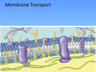
Chapter 5 Membrane Transport Mechanisms. Membrane Permeability. 1) lipid soluble solutes go through faster smaller molecules go faster 1) uncharged & weakly charged go faster 2) Channels or pores may also exist in membrane to allow transport. 1. 2.

E N D
Chapter 5 Membrane TransportMechanisms
Membrane Permeability 1) lipid soluble solutes go through faster • smaller molecules go faster 1) uncharged & weakly charged go faster • 2) Channels or pores may also exist in membrane to allow transport 1 2
How to get other molecules across membranes • There are two ways that the molecules typically move through the membrane: • passive transport and active transport • Active transport requires that the cell use energy that it has obtained from food to move the molecules (or larger particles) through the cell membrane. • Passive transport does not require such an energy expenditure, and occurs spontaneously.
1 Passive Transport • Net movement of material from hi to lo concentration • Diffusion • Osmosis • Facilitated Diffusion concentration gradient
Passive Transport • Simple Diffusion- simple movement from regions of high concentration to low concentration • Osmosis- diffusion of water across a semi-permeable membrane • Facilitated diffusion协助扩散- protein transporters which assist in diffusion
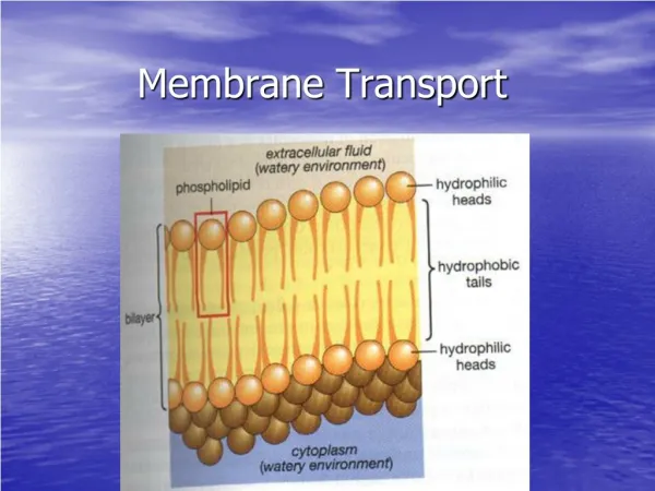
Passive Transport • Unaided movement through the phospholipid bilayer in response to concentration gradients. • Large or charged molecules are unable to pass through the bilayer unassisted.
Diffusion Movement generated by random motion of particles. Caused by internal thermal energy. Movement always from region of high free energy(high concentration) to regions of low free energy (low conc.)
Diffusion • If a concentration gradient exists, there will be a net flow of material across the membrane
Osmosis Movement of water across a semi-permeable barrier. Example: Salt in water, cell membrane is barrier. Salt will NOT move across membrane, water will. Text pg 87
Osmosis • Refers to movement of water • Across a semipermeable membrane • Permeable to water • Impermeable to dissolved materials • Water always moves from lo to high solute • Hypotonic Hypertonic
cell Osmosis in Hypertonic medium Hypertonic solutions- shrink cells Osmosis in Hypotonic medium Hypotonic solutions- swell cells
Osmosis Hypertonic Hypotonic
Plasmolysis due to water deprivation Plant Turgor Hypertonic Hypotonic Central vacuole Water pressure inside cell lends support to plant structure
+ Charged particle cloaked with water molecules, can not get through Na+ Cl- K+ H+
Sugars Large molecules, also cloaked with water molecules, can not get through Nucleotides Amino acids
Sugars Large molecules can not get through Facilitated Diffusion
Transport Protein Shape change resulting from solute interaction with transport protein Channel
Types of Protein Transporters A. Facilitated Diffusion Assist in diffusion process. Solutes go from High conc to Low conc. Examples: Glucose transporters http://bio.winona.msus.edu/berg/ANIMTNS/FacDiff.htm Text pg 88
A Transport ProteinsFacilitated Diffusion & Active Transport • move solutes faster across membrane • highly specific to specific solutes • can be inhibited by drugs
glucose Na+ Transport Proteins are specific
High No transport Cell Controls Movement by Number of Transport Proteins in Membrane Moderate
glucose Gated Channels Chemical messenger, e.g. insulin
Voltage Dependent K+ channel Model of protein shape change in response to change in voltage across a membrane Roderick MacKinnon, M.D., Howard Hughes Medical Institute Investigator, Youxing Jiang, Ph.D.
cysteine cystine Cystinuria Cystine crystal • Defective transport protein in urinary tract unable to absorb cystine from urine
Facilitated DiffusionThe Glucose Transporters • Transport of glucose into cells mediated by proteins in the GLUT (GLUcose Transport) family of transporters. There are 7 different, but related, proteins. But, only four (GLUT1-4) are known to be involved in glucose transport. • All GLUT proteins share a set of similar structural features and are all about 500 amino acids in length (giving them a predicted molecular weight of about 55,000 Daltons) • Glucose uptake shows saturation and glucose uptake can be inhibited by drugs A classic Membrane Transport protein
Glucose TransporterCharacteristics: • integral protein: spans the membrane • 12 alpha helices woven into membrane • 55,000 mol. wt. • Text pg. 88
Glucose Transporter:How it works.. • glucose binds to outside of transporter (exterior side with higher glucose conc.) • glucose binding causes a conform. change in protein • glucose drops off inside cell • protein reassumes 1st configuration
Types of Protein Transporters: Ion Channels • work by facilitated diffusion No E! • deal with small molecules... ions • open pores are “gated”- Can change shape. • How? • How much gets in? • important in cell communication
B Ion Channels • Work fast: No conform. changes needed • Not simple pores in membrane: • specific to different ions (Na, K, Ca...) • gates control opening • Toxins, drugs may affect channels • saxitoxin, tetrodotoxin • cystic fibrosis
Cystic Fibrosis • Fatal genetic disorder • Mucus build-up results in lung and liver failure • Patients die between 4 and 30 yrs. • Single gene defect • 1 in 25 Caucasians carry 1 bad gene copy • 1 in 2500 kids has it in Canada • Testing
CF Cont… • ~Proteins for diffusion of salt into the airways don't work. • ~Less salt in the airways means less water in the airways. • ~ Less water in the airways means mucus layer is very sticky (viscous). • ~Sticky mucus cannot be easily moved to clear particles from the lungs. • ~Sticky mucus traps bacteria and causes more lung infections. http://www.the-aps.org/education/lot/cell/HotT.htm
2 Active Transport • Active transport- proteins which transport against concentration gradient. • Requires energy input T
Types of Protein Transporters: Active Transport • carrier proteins • go against the concentration gradients Low to High • require Energy to function (ATP, PEP, light energy, electron transport)
Membrane Transport Active Transport • Move materials from lo to high concentration • Requires cell to expend energy • Equivalent to running diffusion in reverse • Simulation 1- how can you put the particles back into the box? http://lewis.eeb.uconn.edu/lewishome/applets/Diffusion/diffusion.html
Active Transport Sodium-Potassium Pump CA-ATP Pump proton Pump p- proton Pump v -proton Pump H- ATP Pump Cotransport symport aniport
Active Transport:Sodium-Potassium Pump ATP-direct depend Na+ low Na+ high K+ low K+ high Balance of the two ions goes hand-in-hand ATP required for maintenance of the pump
K+ K+ K+ K+ K+ Na+ Na+ Na+ Na+ Na+ Na+ Na+ Na+ Na+ Na+ Na+ Na+ Na+ Na+ Na+ Electrochemical Gradient negative positive ADP ATP Na+ K+ pump This gradient powers conduction of signals along nerves
sucrose sucrose sucrose sucrose sucrose sucrose sucrose sucrose H+ H+ H+ H+ H+ H+ H+ H+ H+ H+ H+ H+ H+ H+ H+ H+ H+ H+ H+ H+ Stored Energy for Cotransport ADP ATP proton pump Voltage difference or membrane potential
The sodium/potassium pump • All nerve and muscle cells have a high internal potassium ion concentration and a low internal sodium ion concentration. [Ki=166 mM; Ko=5 mM; Nai=18 mM; Nao=135 mM]. • Early on, it was thought that the nerve and muscle membranes were relatively impermeable to these ions and that the difference in ionic concentration was set up in early development of the cells. The membrane then became impermeable. • The later availability and use of radioactive Na and K ions showed that this was not true and that there was a metabolic pump that pumped Na out of the cell and K in; the ratio being 3 Na pumped out of the cell for every 2 K pumped into the cell.
Is a Protein Involved ? • Experiments showed a dependency of both Na and K ions for pump to work • Pump was inhibited by ouabain (a drug) • 1957: an ATPase enzyme was found to be associated with Na/K pumping • Studies showed this ATPase capable of pumping Na/K ions
Sodium/PotassiumATPase Protein • Made of 2 large and 2 small subunits • 2 large units span membrane • inside region: contains ATP binding site • inside: binding sites for Na • outside: binding site for K • How does it work??