252 Pre-lab lecture
252 Pre-lab lecture. Human Renal Function “The Pee lab”. Nephron. The nephron is the functional unit of the kidney. Many filtered substances are reabsorbed into the blood. Certain substances are secreted from the blood into the tubule to become excreted. Nephron anatomy (page1025).

252 Pre-lab lecture
E N D
Presentation Transcript
252 Pre-lab lecture Human Renal Function “The Pee lab”
Nephron • The nephron is the functional unit of the kidney. • Many filtered substances are reabsorbed into the blood. • Certain substances are secreted from the blood into the tubule to become excreted.
Nephron anatomy (page1025) • Filtrate passes from proximal convoluted tubule to descending loop of Henle to ascending loop of Henle to distal convoluted tubule to the collecting duct.
Reabsorption • PCT: 65% water, Na and K. 100% glucose and amino acids. 50% CL. 80-90% filtered HCO3. 50% filtered urea. • LOH: 15% of the filtered water. 20-30% of Na and K. 35% of filtered Cl. 10-20% of HCO3. • Early DCT: 10-15% water and trace ions • Late DCT: depends on the body’s needs.
Most solute reabsorption involves Na + ions. • About 90% of water reabsorption is termed obligatory because water is “obliged” to follow solutes.
Paracellular vs Transcellular Reabsorption • Paracellular reabsorption is a passive process where substances leak between tubular cells • Transcellular reabsorption is a process where substances pass from the tubular lumen, through the apical membrane of a tubular cell, across the cytosol and through the basolateral membrane.
Primary vs secondary active transport • Primary active transport requires ATP and pupms substances across a membrane • Secondary active transport uses energy stored in an electrochemical gradient to move substances across a membrane.
Glucose reabsorption (page 1037) • Na+/glucose symporter: glucose follows Na+ across the apical membrane through the symporter . • This process occurs due to a Na + concentration gradient established by the Na+/K-pump. • Glucose moves across the basolateral membrane by facilitated diffusion. • Na + and glucose diffuse into the capillary.
Reabsorption of Na+ and HCO3-and secretion of H+ • Na+/H+antiporter simultaneously moves Na+ into the cell (down conc gradient) and H+ ions into the tubule (secretion). • Na+ conc gradient established by NA+/K-pump. • HCO3- move through basolateral membrane via facilitated diffusion. • NA +and HCO3- diffuse into capillary.
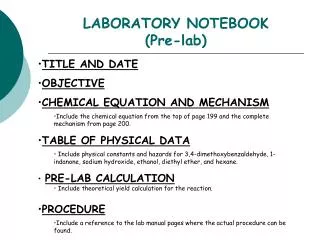
252 Pre-lab lecture Chemical Aspects of Digestion
4 exercises Digestion of protein Digestion of Lipids Digestion of Starch Blood glucose concentration
Blood glucose regulation Glucagon is pancreatic hormone, which accelerates both the conversion of glycogen(storage form) to glucose and a process called gluconeogenesis. Insulin is a pancreatic hormone, which accelerates both the facilitated diffusion of glucose into cells and also the conversion of glucose into glycogen. Normal blood glucose is 80 to 110 mg/dl or 4 to 6 mmol/l
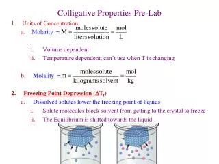
Post-meal condition Hepatic vein 100mg/dl Small intestine liver Hepatic portal vein (120mg/dl Superior mesenteric artery (90mg/dl)
Post-meal condition Superior mesenteric artery is low in glucose. Hepatic portal vein is high in glucose (absorption from the small intestine). Insulin causes the liver to uptake glucose… Hepatic vein maintains normal blood glucose.
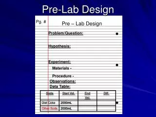
Pre-meal condition Hepatic vein 100mg/dl Small intestine liver Hepatic portal vein (80mg/dl Superior mesenteric artery (80mg/dl)
Pre-meal condition Superior mesenteric artery is still low in glucose. Hepatic portal vein is low in glucose. Glucagon causes the liver to convert glycogen to glucose and thus glucose is released into the bloodstream. Hepatic vein maintains normal blood glucose.
Digestion This is the process of breaking down large molecules into smaller molecules that can be absorbed into the blood. Digestive enzymes catalyze the breakdown of these molecules: Salivary Amalyse (starch to maltose) Pepsin (protein to polypeptides) Pancreatic Lipase (lipids to glycerol+ 3 fatty acids)
Testing for digestion Digestive enzyme activity is affected by factors such as temperature, ph, surface area and time. Testing for digestion usually involves testing for the presence of products.
Female Reproductive Cycle • Controlled by monthly hormone cycle of anterior pituitary, hypothalamus & ovary • Monthly cycle of changes in ovary and uterus • Ovarian cycle • changes in ovary during & after maturation of oocyte • Uterine cycle • preparation of uterus to receive fertilized ovum • if implantation does not occur, the stratum functionalis is shed during menstruation
Hormonal Regulation of Reproductive Cycle • GnRH secreted by the hypothalamus controls the female reproductive cycle • It stimulates the anterior pituitary to secrete FSH & LH • FSH initiates growth of follicles that secrete estrogen • estrogen maintains reproductive organs • LH stimulates ovulation & promotes formation of the corpus luteum which secretes progesterone and estrogen. • progesterone prepares uterus for implantation and the mammary glands for milk secretion • In males LH causes the Interstitial cells to produce testosterone.
Menstrual Phase • Menstruation lasts for 5 days • First day is considered beginning of 28 day cycle • In ovary • 20 follicles that began to develop 6 days before are now beginning to secrete estrogen • In uterus • declining levels of progesterone caused uterine arteries to constrict -- glandular tissue dies • stratum functionalis layer is sloughed off along with 50 to 150 ml of blood
Preovulatory Phase • Lasts from day 6 to 13 (most variable timeline) • In the ovary (follicular phase) • Secretion of FSH is slowing. • dominant follicles survives to day 6 • by day 14, graafian follicle has enlarged & bulges at surface • Increasing estrogen levels trigger the secretion of LH • In the uterus (proliferative phase) • increasing estrogen levels have repaired & thickened the stratum functionalis to 4-10 mm in thickness
Ovulation • Rupture of follicle & release of 2nd oocyte on day 14 • Cause • increasing levels of estrogen stimulate release of GnRH which stimulates anterior pituitary to release more LH (spike)
Postovulatory Phase • Most constant timeline = lasts 14 days • In the ovary (luteal phase) • No Fertilization…. corpus luteum regresses • as hormone levels drop, secretion of GnRH, FSH & LH rise. • Fertilization… developing embryo secretes human chorionic gonadotropin (hCG) which maintains health of corpus luteum & its hormone secretions • In the uterus (secretory phase) • hormones from corpus luteum promote thickening of endometrium to 12-18 mm • formation of more endometrial glands & vascularization • if no fertilization occurs, menstrual phase will begin