Download
1 / 87
880 likes | 1.22k Vues
Shoulder 101 …and Then Some. Evan D. Ellis MD Rebound Orthopaedics and Sports Medicine. Why Shoulder 101?. Multiple studies: High percentage of visits to see PCP are for musculoskeletal pain 2 studies * : Large gap in PCP confidence in evaluating and treating musculoskeletal injuries

E N D
Shoulder 101 …and Then Some Evan D. Ellis MD Rebound Orthopaedics and Sports Medicine
Why Shoulder 101? • Multiple studies: High percentage of visits to see PCP are for musculoskeletal pain • 2 studies*: Large gap in PCP confidence in evaluating and treating musculoskeletal injuries • Studies in both a rural and tertiary academic setting *Lynch et al JBJS AM 2006 and AJO 2005
The Shoulder • ANATOMY • HISTORY • PHYSICAL EXAM • IMAGING • CASES/TREATMENT
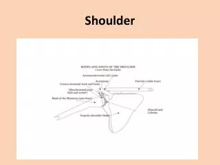
Anatomy • Not a ball and socket • More of a ball on a dish • Static Restraints • Dynamic Restraints
Anatomy • Glenoid Concavity: • Bone • Cartilage • Labrum
Anatomy • Labrum: • Deepens glenoid by 50% • 9 mm superoinferior* • 5 mm anteroposterior* • Contributes to 20% of stability in A-P direction • Loss of labral integrity may result in instability *McMahon et al. JSES. 2004. Jan-Feb;13(1):39-44. *Howell SM, Galinat BJ. The glenoid-labral socket: a constrained articular surface. Clin Orthop. 1989
Static Restraints Glenohumeral Ligaments Superior: Prevents inferior translation with arm at side Middle: Important for mid-range abduction Inferior: Critical for ABD/ER Anterior band prevents anterior inferior translation Anatomy
Anatomy • Ligaments do not center the head. • Limit its translation and rotation. • Think Check-Rains
Anatomy Dynamic Restraints • Muscular Stabilizers • Anterior: Subscapularis • Superior: Supraspinatus • Posterior: Teres minor and Infraspinatus • Lateral: Deltoid • Scapular stabilizers
History Basics • Painful shoulders can be: • Unstable • Stiff • Weak • Rough/Pain “What bothers you about your shoulder?”
History • Age • Gender • Was there an injury? • Injury mechanism • Prior problem • Dominant arm
History • Chronicity • Location of Pain • Pain at night • Stiffness/Unstable • Prior treatment
Physical Exam • Goal: Reproduce Symptoms • Inspection, Palpation, ROM, neurovascular exam, special tests • Compare to contralateral side • Cervical spine • Note provocative positions
Physical Exam • EXPOSE: • Neck • Shoulders • Arms
Physical Exam • EXPOSE: • Neck • Shoulders • Arms • Women need gown or tank top!
Physical Exam Motion: Active/Passive • Forward Elevation • External Rotation • ER in Abduction • Internal Rotation • IR in Abduction • X-Body
Range of Motion • FE: 180 • ERS: 60 • ERA: 90 • IRA: 70 • IRB: T-spine • X-Body: 60
Range of Motion • FE: 180 • ERS: 60 • ERA: 90 • IRA: 70 • IRB: T-spine • X-Body: 60
Range of Motion • FE: 180 • ERS: 60 • ERA: 90 • IRA: 70 • IRB: T-spine • X-Body: 60
Range of Motion • FE: 180 • ERS: 60 • ERA: 90 • IRA: 70 • IRB: T-spine • X-Body: 60
Range of Motion • FE: 180 • ERS: 60 • ERA: 90 • IRA: 70 • IRB: T-spine • X-Body: 60
Range of Motion • FE: 180 • ERS: 60 • ERA: 90 • IRA: 70 • IRB: T-spine • X-Body: 60
Rotator Cuff Exam • MOTOR • Subscapularis • Supraspinatus • Infraspinatus • Teres Minor
Rotator Cuff Exam • MOTOR • Subscapularis • Supraspinatus • Infraspinatus • Teres Minor
Rotator Cuff Exam • MOTOR • Subscapularis • Supraspinatus • Infraspinatus • Teres Minor
Rotator Cuff Exam • MOTOR • Subscapularis • Supraspinatus • Infraspinatus • Teres Minor
Rotator Cuff Exam • MOTOR • Subscapularis • Supraspinatus • Infraspinatus • Teres Minor
Neurologic Exam • NEURO • Sensation • Motor • Reflexes • Spurling’s
Neurologic Exam • NEURO • Sensation • Motor • Reflexes • Spurling’s
Special Tests - Cuff • CUFF • Neer Impingement Sign • Neer Impingement Test • Subacromial injection • Hawkins Test
Special Tests - Cuff • CUFF • Neer Impingement Sign • Neer Impingement Test • Subacromial injection • Hawkins Test
Special Tests - Cuff • CUFF • Neer Impingement Sign • Neer Impingement Test • Subacromial injection • Hawkins Test
Apprehension/Relocation Supine position Stabilizes scapula Abduct to 90° Increase ER gradually Positive: Apprehension w/ increasing amounts of ER Apprehension relieved by posterior force on the humerus Special Tests - Instability
Seated Load & Shift Assess A & P translation Grade 1+: to rim 2+: over rim w/reduction 3+: over rim & locked Compare to other side Assess for pain, click, & instability Special Tests - Instability
Supine Load & Shift Arm position: 45-60° abduction Ant/Post directed force applied to humerus Assess Stability Pain Palpable click Special Tests - Instability
Sulcus Sign: Arm at side To look for multi-directional instability Grade 1+ = 1 cm 2+ = 1-2 cm 3+ = > 2 cm Look for generalized hypermobility Special Tests - Instability
Radiographs • Never order an MRI before X-Rays • Everyone deserves a normal set of X-Rays! • Most important X-Rays: True AP (Grashey) and Axillary Lateral • These two X-Rays are almost always omitted from a “shoulder series”!
Radiographs • True AP or Grashey View: • Arthritis • Fracture • Massive Rotator Cuff tear
Radiographs • True AP
Radiographs • Axillary Lateral • Arthritis • Instability • Fracture
Radiographs • Axillary Lateral
Radiographs • Additional Views • Outlet • Internal/External • Stryker Notch • West Point View
The Shoulder • Diagnosable & Treatable • Rotator cuff tears • Shoulder instability • Arthritis • SLAP tear • Diagnosable & Untreatable • Brachial neuritis • Voluntary instability/MDI • Rib fractures
Case #1 History: • 16 year old RHD male football player • Shoulder “popped” out of place while getting tackled • To ER for reduction • Has happened 2 previous times
Case #1 Physical Exam: • Full Range of Motion • Full rotator cuff strength • + Apprehension Test • + Relocation Test • + Anterior Load & Shift
Case #1 • What do you do? • Place him in a sling • Refer to Ortho • If first time dislocater – Physical Therapy • If 2 or more dislocations – MRI and surgery
Regular Sling vs. External Rotation Which is better? Itoi, JBJS 2007 159 patients Avg follow up of 25.6 months 74 immobilized in IR 31 recurred (42%) 85 immobilized in ER 22 recurred (26%) *Effect on labral position for healing Sling