Download
1 / 35
360 likes | 748 Vues
Tissues, Organ Systems and Homeostasis. B. L. Krilowicz Biology 155 Spring 2010. Organization of the Animal Body. Animals’ bodies exhibit hierarchical organization – Biological molecules are organized into organelles (ex. Phospholipids and proteins are arranged into the plasma membrane)

E N D
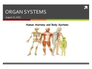
Tissues, Organ Systems and Homeostasis B. L. Krilowicz Biology 155 Spring 2010
Organization of the Animal Body • Animals’ bodies exhibit hierarchical organization – • Biological molecules are organized into organelles (ex. Phospholipids and proteins are arranged into the plasma membrane) • Organelles are organized into a cell (ex. Nucleus + plasma membrane + cytoplasm proper + many organelles = cell)
Organization of the Animal Body (continued) • Hierarchical organization continued – • Groups of similar cells are organized into tissues (ex. Cardiac muscle cells are organized into the tissue, cardiac muscle); Note that the evolution of multicellular living forms required development of tissues • Two or more tissues are organized to form an organ (ex. Cardiac muscle tissue + connective tissue + epithelial tissue = the heart; provides force to move blood)
Organization of the Animal Body (continued) • Hierarchical organization continued – • Organs are organized into organ systems (ex. Heart + blood vessels + blood = cardiovascular system; function is transport) • Organ systems are organized into an organism (ex. An animal consists of 11 organ systems)
Embryonic Tissues – all adult tissues are derived from one of three embryonic tissues Ectoderm = “outside skin” gut Mesoderm = “middle skin” Cross section through embryo Endoderm = “inside skin” Animal embryo
Fate of Embryonic Tissues • Ectoderm will become the outer covering of the body and the nervous system • Mesoderm will become the muscles and internal skeletons • Endoderm will become the lining of the gastrointestinal tract, lungs, vessels and ducts
Adult Tissues • Definition = groups of cells with similar structure, embryonic origin, and function; cells are bound together by extracellular material and function together to perform a specific task • There are four main types of adult tissues in the animal body
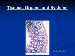
Epithelial Tissues • Source = may be derived from any tissue in the embryo • Function = mainly protective, therefore they cover all free surfaces of the body; can be specialized for absorption, excretion, secretion, etc.
Epithelial Tissues (continued) • Characteristics = • Closely joined cells with little extracellular material between the cells • Presence of a basement membrane secreted by the epithelial cells; separates the epithelial cells from underlying tissues • One free surface not in contact with other cells
Epithelial Tissues (continued) • Classification = ask and answer two questions • How many cell layers above the basement membrane? • Simple = one layer of cells; used for exchange (ex. Diffusion of gases in the alveoli of the lungs, absorption in the small intestine; see previous slide) • Stratified = more than one layer of cells; used for protection (ex. the outer layer of the skin; see board)
Epithelial Tissues (continued) • Classification continued – • What shape are the cells? (when viewed from the side) • Flat and thin = squamous; used to maximize diffusion (non-energy requiring exchange where things move from an area of high to an area of low concentration); ex. Alveoli of lungs, capillaries • Look like squares = cuboidal; used to maximize energy requiring exchange such as absorption and excretion (things can move from an area of low concentration to an area of high concentration); ex. respiratory system • Tall and thin = columnar; as cuboidal above; ex. Small intestine
Connective Tissues • Source = may be derived from any tissue in the embryo • Function = many, but generally holds things together in the body; can be specialized to give structure to and protect body parts
Connective Tissues (continued) • Characteristics = • Few cells • Lots of extracellular material between the cells; extracellular material is produced by the cells and is called matrix • Matrix consists of – • Protein fibers • Ground substance = non-fibrous proteins + other molecules • Fluid
Connective Tissues (continued) • Classification = ask and answer one question • What is the nature of the extracellular matrix? • Fluid = tissue is blood, functions in gas transport
Connective Tissues (continued) • Classification = ask and answer one question • What is the nature of the extracellular matrix? (continued) • Solid = • Mainly protein fibers = connective tissue proper (ex. Loose and dense connective tissues, adipose) • Protein fibers + ground substance = special connective tissues (ex. Bone and cartilage)
Muscle Tissues • Source = derived from the embryonic mesoderm • Function = allows movement of the body or movement within the body • Characteristics = • Closely joined cells with little extracellular material • Contain specialized protein fibers capable of contraction
Muscle Tissue (continued) • Classification – • Cardiac Muscle = heart muscle • One, centrally located nucleus • Presence of striations • Short, branched cells • Presence of intercalated discs • “involuntary”
Muscle Tissue (continued) • Classification – • Skeletal Muscle • Many, peripherally located nuclei • Presence of striations • long, thin cells • “voluntary”
Muscle Tissue (continued) • Classification – • Smooth Muscle – found in “hollow organs” • One, centrally located nucleus • no striations • Short, tapered cells • “involuntary”
Nervous Tissue • Source = derived from the embryonic ectoderm • Function = communication • Characteristics – • Electrically excitable cells (neurons with cell body and processes), or • Cells that support, nourish and protect the neurons (glia) • Classification = none
Fig. 20.10 Animal Organ Systems
Fig. 20.10 Animal Organ Systems - continued
Fig. 20.10 Animal Organ Systems - continued
Fig. 20.10 Animal Organ Systems - continued