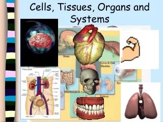
Tissues and Organ Systems
760 likes | 965 Vues
Tissues and Organ Systems. There are between 75 and 100 trillion cells in the body of an adult human They are programmed to form different groups that have specific functions; this occurs due to: The contents of the cell’s cytoplasm --> how organelles are distributed

Tissues and Organ Systems
E N D
Presentation Transcript
Tissues and Organ Systems • There are between 75 and 100 trillion cells in the body of an adult human • They are programmed to form different groups that have specific functions; this occurs due to: • The contents of the cell’s cytoplasm --> how organelles are distributed • Environmental conditions, such as temperature--> affects the activity of certain molecules in the cell • The influence of neighbouring cells--> Surrounding cells can send chemical messages that affect how cells around it will develop
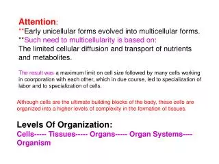
Cell Specialization and Development • Cells become differentiated because of genes that get turned off. • They only express a certain set of information. • Environmental factors (hazardous chemicals, smoking) can affect the expression of these genes leading to abnormalities
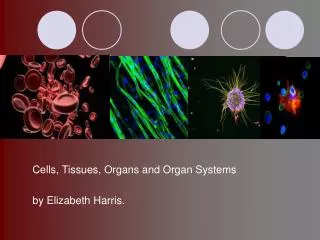
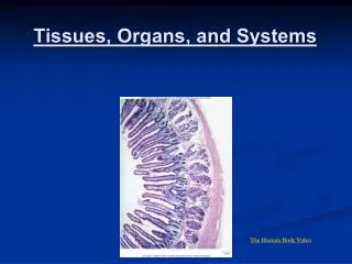
Types of Tissues • All animals have four main types of tissues • Epithelial: line the surfaces of the body, both as a body covering and between internal organs; form a barrier • Muscle: designed to change their shape by shortening or lengthening • Nervous: neurons receive and transfer signals to coordinate body actions • Connective: strengthens, supports, protects, binds, or connects cells and tissues.
Epithelial tissue • Skin epithelia: made of thin, flat cells that form sheets and act as a semi-permeable barrier between the inside and outside of a body • Columnar Epithelia: made of columns of cells that line the small intestine, stomach and glands; may secrete mucus and have finger-like projections, and absorb materials
Muscle • Skeletal muscle: cells line up in the same direction gives it a striped/striated look; attaches to bone making it possible to move • Smooth muscle: cells are tapered at both ends, found in blood vessels and walls of internal organs, they contract more slowly than skeletal muscle • Cardiac muscle: found only in the heart, contracts as a unit
Nervous Tissue • Have various actions: • Some relay signals from the brain or spinal cord to muscles and glands • Others detect information form their environment and trigger the body’s responses
Connective Tissue • Bone: made of cells surrounded by calcium-hardened tissue through which blood vessels run, needed for movement, support and protection • Fat (adipose tissue): made of large, tightly packed cells found under the skin and around organs; needed for energy storage, padding and insulation • Blood: includes red blood cells (RBCs), white blood cells (WBCs) and platelets within a liquid called plasma; used to transport nutrients and oxygen, clots when injury occurs, attacks foreign cells such as bacteria and viruses
Stem Cells • Just as plants have meristematic cells that can give rise to different plant tissues, animals have stem cells that can divide and will eventually will rise to specialized and differentiated tissues. • A stem cell is an unspecialized cell that can produce various specialized cells • These stem cells are abundant in an embryo 24 hours after fertilization. They are called totipotent because they can become any kind of cell in the body. • At 5 days the stem cells are called pluripotent and are capable of producing many but not all kinds of cells
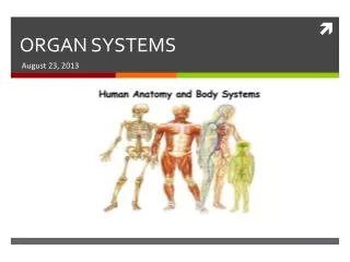
Organs and Systems • Specialized tissues formed from stem cells work together to form organs which work together to perform complementary functions to form organ systems • Organ systems can be observed using common medical imaging technologies that allow physicians to diagnose disorders
Medical Imaging Technologies • X Ray: electromagnetic radiation is transmitted through the body to expose photographic film on the other side • CT/CAT scan: reconstruction of X rays of thin slices of a body part using a computer • Ultrasound sonography: produced by directing high frequency sound waves at a part of the body that shows real-time movement of body parts • MRI scan (magnetic resonance imaging): produced using radio signals in a magnetic field to create images of body parts
Hierarchical Organization of the Body Atom molecules organelles cells tissues organs organ system organism * Four main types of tissues: connective, epithelial, nervous & muscle.
Digestive System Overview • Known as gastrointestinal (GI) tract or alimentary canal. • Open at both ends to the outside world. • Consists of long convoluted tube with accessory organs. • Components: mouth, pharynx, esophagus, stomach, small intestine, large intestine & anus. • Accessory organs include: salivary glands, pancreas, liver & gallbladder.
Steps in Digestion • Ingestion • Digestion • Absorption • Elimination
Types of Digestion Mechanical • Food mass is shredded, torn & churned. • Occurs in mouth & stomach. Chemical • Chemicals and enzymes break down food. • Occurs in mouth, stomach & small intestine.
Organs of the Digestive System • Oral Cavity • Esophagus • Stomach • Small Intestine • Large Intestine
Oral Cavity • Lips, tongue, teeth & jaw muscles break food down into smaller pieces. • Food mixes with saliva and amylase, which begins the chemical digestion of carbohydrates. • A bolus (moistened ball-like mass) forms and is swallowed. • The bolus passes through the pharynx, past the epiglottis & through the esophagus.
Esophagus • Muscular tube that connects pharynx & stomach. • The cardiac or lower esophageal sphincter connects the esophagus to the stomach. • Constriction of this ring of smooth muscle prevents reflux (ensures one-way flow of food). • Peristalsis is a series of coordinated muscular contractions that propels food along the digestive tract.
Stomach • J-shaped stretchable organ. • Acts as a reservoir for food (1.5 L). • Two sphincters control the movement of food coming into and out of the stomach. • Cardiac sphincter: Between the stomach & the esophagus. • Pyloric sphincter: Between the stomach & the small intestine (the duodenum).
Stomach • Smooth muscle forms folds (rugae) that allow the stomach to expand. • Mechanical digestion: Walls churn & squeeze bolus. • Chemical digestion: Bolus mixes with gastric juices. • Hydrochloric acid secreted by gastric glands. • Pepsinogen released & converts to pepsin (enzyme that breaks down proteins). • Bolus becomes a liquefied paste (chyme).
Stomach • Mucus cells secrete mucus to line & protect stomach from HCl (aq). • Ulcers: HCl (aq) burns a hole through the mucus, irritating the stomach cells below • Usually the result of a particular type of bacteria (antibiotics required).
Small Intestine • Major site of digestion & absorption (80% occurs). • About 6 m long & has a SMALLER diameter than the large intestine. • Lined with tiny finger-like projections called villi, which project into the lumen. • Microvilli line the villi. • Villi & microvilli increase the surface area for absorption.
Small Intestine • Consists of three sections: • Duodenum • Jejunum • Ileum.
Small Intestine • Mechanical digestion: Alternating contraction & relaxation of the smooth muscle mixes chyme with intestinal juices & secretions from the pancreas & liver. • Nutrients absorbed into capillaries in the villi. • Nutrients transported to the liver & then to all body cells. • Products from fat digestion are absorbed into lacteals, which connect to the lymphatic system.
Large Intestine • Reabsorbs water, salt & some vitamins. • Holds & compacts unabsorbed material. • About 1.5 m long & has a LARGER diameter than the small intestine. • Consists of four sections: • Caecum • Colon • Rectum • Anus.
Large Intestine • Chyme passes from the small intestine into the caecum, through the ileocaecal valve. • Waste products accumulate & are compacted into feces (3/4 water, 1/4 solid matter) • Feces pass through the rectum & exit the body through the anus. • Appendix attached to caecum, exact function is unknown.
Large Intestine • Defecation controlled by two sphincters: • Rectal sphincter: Between the large intestine & the rectum. • Anal sphincter: Between the rectum & the anus.
The Circulatory System • The functions of the circulatory system is to pick up and transport nutrients and oxygen to cells and carries wastes to the organs responsible for eliminating them from the body.
Circulation in Various Organisms • Insects have a circulatory system that is ‘open.’ They have one major vessel that empties oxygenated blood into parts of the body. Cells are literally bathed in blood. • Mammals and birds have a ‘closed’ circulatory system - the blood stays within tubes or vessels. • Fish have two-chambered hearts, amphibians have three-chambered hearts.
The 3 main parts of TheCirculatory system • The Heart • The Blood Vessels • Blood
Parts of The Heart • The Atria -Receiving Chambers • The Ventricles -Pumping Chambers • The Valves -Controls Flow • The Septum -Divides the Heart
Types of Blood Vessels • Arteries -Carry blood away from the Heart -The Aorta is the largest artery • Veins -Carry blood away from the Heart -Veins contain valves -The Vena Cava is the largest vein • Capillaries -Known as the “Distribution Pipes”
Passage of Blood Through the Heart • Blood follows this sequence through the heart: superior and inferior vena cava → right atrium → tricuspid valve → right ventricle → pulmonary semilunar valve → pulmonary trunk and arteries to the lungs → pulmonary veins leaving the lungs → left atrium → bicuspid valve → left ventricle → aortic semilunar valve → aorta → to the body. • N.B. The pulmonary artery is the only artery in the body that carries deoxygenated blood. • The Pulmonary vein is the only vein that carries oxygenated blood. http://www.bishopstopford.com/faculties/science/arthur/Heart%20drag&drop.swf
The pumping of the heart sends out blood under pressure to the arteries. Blood pressure is greatest in the aorta; the wall of the left ventricle is thicker than that of the right ventricle and pumps blood to the entire body. Blood pressure then decreases as the cross-sectional area of arteries and then arterioles increases.
Path of blood through the heart Blood Flow through the Human Heart Blood circulation
The Composition of Blood • The Plasma (Fluid) makes up 55% of the blood volume. • The Solids (Cells) make up 45% of the blood volume.
Blood Plasma • 97% Water • Other 3% -Antibodies and Proteins - Nutrients and Wastes
Blood Solids • Red Blood Cells -Carry oxygen -Contain Hemoglobin • White Blood Cells -Attack bacteria & other invaders • Platelets -Control the blood clotting process
Blood Solids • Red Blood Cells -Carry oxygen -Contain Hemoglobin • White Blood Cells -Attack bacteria & other invaders • Platelets -Control the blood clotting process
Cardiovascular Disorders • Cardiovascular disease (CVD) is the leading cause of death in Western countries. • Modern research efforts have improved diagnosis, treatment, and prevention. • Major cardiovascular disorders include atherosclerosis, stroke, heart attack, aneurysm, and hypertension.
Atherosclerosis • Atherosclerosis is due to a build-up of fatty material (plaque), mainly cholesterol, under the inner lining of arteries. • The plaque can cause a thrombus (blood clot) to form. • The thrombus can dislodge as an embolus and lead to thromboembolism. Atherosclerosis Calcification
Stroke, Heart Attack, and Aneurysm • A cerebrovascular accident, or stroke, results when an embolus lodges in a cerebral blood vessel or a cerebral blood vessel bursts; a portion of the brain dies due to lack of oxygen. • A myocardial infarction, or heart attack, occurs when a portion of heart muscle dies due to lack of oxygen.
Coronary Bypass Operations • A coronary bypass operation involves removing a segment of another blood vessel and replacing a clogged coronary artery. • It may be possible to replace this surgery with gene therapy that stimulates new blood vessels to grow where the heart needs more blood flow.