Download
1 / 22
220 likes | 465 Vues
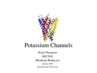
Calcium-Activated Potassium Channels: Multiple Contributions to Neuronal Function. E.S. Louise Faber and Pankaj Sah PPT by Anvinh Nguyen. Calcium-activated Potassium Channels. Found throughout CNS Activated by rises in cytosolic calcium during AP

E N D
Calcium-Activated Potassium Channels: Multiple Contributions to Neuronal Function E.S. Louise Faber and PankajSah PPT by Anvinh Nguyen
Calcium-activated Potassium Channels • Found throughout CNS • Activated by rises in cytosolic calciumduring AP • These channels are targets for modulation because they have been thought to influence neurological and psychiatric disorders.
First Identification of calcium-dependent potassium channels… • By Gardos– he worked with red blood cells. • By Meech and Strumwasser – they described an ionic current activated by a rise in cytosolic calcium.
Three Types of Calcium-activated Potassium Channels • 1. BK Channels • First type identified and cloned • Highly potassium selective and large conductance • 2. SK Channels • Fairly low conductance • 3. IK Channels • Intermediate conductance
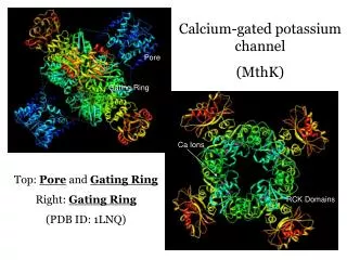
BK Channels • Large conductances 200 to 400 pS • Requires both intracellular calcium and depolarization • Surprisingly, can open without calcium calcium and voltage are independent processes • α (tetramer, pore-forming) and β (three subunits) subunits
BK Channel Enhancement and Inhibition • BK Channels are enhanced by Dehydrosoyasaponin-1 (DHS-1) • BK Channels are inhibited by TEA
SK Channels • Three types have been successfully cloned: SK1, SK2, and SK3 • Conductance of 2 to 20 pS • Activated by intracellular calcium, and are voltage insensitive • SK pore is similar to the voltage-gated potassium channel’s pore • Must both be transmembrane bound proteins with tetramer pore
SK Channel Enhancement and Inhibition • SK Channels are enhanced by 1-ethyl-2-benzimidazolinone (EBIO) • It works by changing calcium sensitivity and open probability • SK Channels are inhibited by Apamin, a type of bee venom
IK Channels • Conductance of 20 to 100 pS • Identified in epithelial and red blood cells • Activated by intracellular calcium, and are voltage insensitive
IK Channel Enhancement and Inhibition • IK Channels are also enhanced by 1-ethyl-2-benzimidazolinone (EBIO) • IK Channels are inhibited by certain neurotransmitters
Afterhyperpolarization (AHP) • AHP follows an action potential in many neurons • They can last up to several seconds • All cases: slow component AHP from calcium-activated potassium conductance • Some cases: intracellular calcium release helps activate AHP
Types of AHPs • Fast: • 1. Fast • Slow: • 2. Medium • 3. Slow • Fast AHP responsible for: • Repolarization • Slow (medium and slow) AHP responsible for: • limiting firing frequency • Generating spike-frequency adaptation
Fast AHP • Mediate by calcium-activated potassium currents • Calcium and depolarization • Blocked by TEA, so BK channel is most likely the one • Modulated by PKA through phosphorylation • Upreg or downreg is based on the channel
Fast AHP Introduction of paxilline (could also be TEA) would cause a reduction in the fast AHP.
Medium AHP • Mediate by calcium-activated potassium currents • Calcium only • It is blocked by apamin, not TEA, suggesting SK channel • Expression of SK1, SK2, and SK3 resulted in the same result as the medium AHP • Does not influence repolarization like the fast AHP.
Medium AHP Apamin blocks SK Channels which causes a reduction in the medium AHP. This causes: Higher firing frequency Decrease in spike frequency adaptation In figure A, slow AHP is still there, only medium AHP was reduced
Slow AHP • More commonly seen in AP trains than single AP’s • Slow AHP is not blocked by either TEA or apamin • Slow AHP is modulated by a range of neurotransmitters IK channel • Monoamines can activate PKA or inhibit calcium-induced calcium release. • It is responsible for spike frequency adaptation
Slow AHP Adding noradrenaline causes the slow AHP to be reduced. This also resulted in a reduction in spike frequency adaptation.
Functional Role: Medium AHP • Blockade of SK channels with apamin learning in a number of behavioral studies • Effects the acquisition of learning the task, not the consolidation
Functional Role: Slow AHP • Reduction in the Slow AHP reduction in spike frequency adaptation • Inhibition of Slow AHP increased excitability during learning and neuronal plasticity
Aging • Drugs that depress the slow AHP, such as calcium channel blockers have shown to effectively improve learning in aged animals. • Further studies show that an increase in medium AHP reduced ability of hippocampal neurons from aged animals to undergo synaptic plasticity.
Disease • Apamin binding sites reduced in hippocampus brains from patients with Alzheimer’s disease • Slow AHP has been proposed to reduce excitability to prevent onset of epilepsy in CA1 hippocampal neurons.