Download

1 / 58
580 likes | 878 Vues
William 2001. Intracranial hemorrhage and brain disorders. Intraventricular hemorrhage Brain disorders Cerebral palsy Neonatal encephalopathy . Types of intracranial Hg: Subdural Subarachnoid Intracerebellar Periventricural - intraventricural : -- In term infants:

E N D
William 2001 Intracranial hemorrhage and brain disorders
Intraventricular hemorrhage • Brain disorders • Cerebral palsy • Neonatal encephalopathy
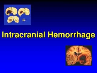
Types of intracranial Hg: • Subdural • Subarachnoid • Intracerebellar • Periventricural - intraventricural: -- In term infants: ½ due to trauma/asphyxia ¼ due to unknown causes intraventricular Hemorrhage
-- In preterm infants: Due to multifactorial factors: • Hypoxic - ischemic • Anatomical causes • Coagulopathy Periventricular – intraventricular Hg: - Fragile capillaries in germinal matrix rupture Hg
- May extend to the ventricles or brain parenchyma - Common in neonates < 34 weeks but may occur in older infants - Starts usually within 72 hours but may develop 24 days after birth - External perinatal and postnatal factors may alter it’s % and severity
- Minimal in ½ of the cases no C/P - Mostly small Hg or Hg confined to the ventricles resolve without neurological impairment - In large Hg hydrocephalus or periventricularleukomalacia CP Pathology: - Due to damage of germinal matrix
capillary network - ↑ in preterm infants due to: • Poor support • Venous anatomy in this area stasis Hg • Vascular autoregulation is impaired < 32 weeks
Extensive Hg death or handicap due to periventricularleukomalacia: = Cystic area due to ischemia or Hg Incidence and severity: = 4% in term infants = ½ infants < 32 weeks are born withsome Hg minimal effect
Very LBW have the: • Earliest onset • Greatest progression • Highest mortality rate Assessed by: U/S and CT
Grades: I matrix Hg II intraventricular Hg III dilatation of the ventricles IV parenchymal Hg Survival: > 90% in I & II -- 3.2% handicap 50% in III & IV
Very LBW infants: 45% intraventricular Hg 20% of them are III & IV degree Contributing factors: - Prematurity and it’s complications: • Infection ischemia • Acidosis X 3 ↑ risk for grade III & IV if pH < 7.2 • RD and mechanical ventilation
Heparin X 4 Hg - Postnatal factors: • RD • Ventilation therapy • PCO2≥ 60 mmHg within 1st 2 hours • PO2< 40 mmHg within 1st 2 hours • Pneumothorax
Treatment: 1 - Antenatal corticosteroids: ↓ mortality ↓ RD ↓ intraventricular Hg + benefit in cases of PROM Betamethasone ↓ leukomalacia compared to dexamethasone
2 – Phenobarbital and vit K = controversial 3 - Vit E ↓ severity and % but does not ↓ mortality 4 - Indomethacin ↑ mortality in infants < 1000 gm 5 - MgSO4 ↓ periventricular Hg
Prevention: • Avoid hypoxia • CS in preterm cephalic fetus no evidence Studies: - No significant difference - ↓ early intracranial Hg
Outcome in extreme prematurity: • ↑ mortality • ↑ neurological injury • ↑ ophthalmological injury • ↑ pulmonary injury Age α 1 / severe neurological abnormalities
1862 = abnormal labor spasticity 1900 = Sigmund Freud many abnormalities can spastic rigidity Cerebral palsy is caused by a combination of: • Genetic factors • Environmental factors • Physiological factors • Obstetric factors Brain disorders
Still many doctors are afraid of CP from obstetric factors ↑ CS to 1 : 4 births in US with no ↓ in CP
Asphyxia: • Profound metabolic or mixed acidemia < 7 • Persistent Apgar score 0 – 3 for > 5 min • Neurological sequelae: - Seizures - Coma - Hypotonia - Dysfunction of ≥ 1 system : GIT - Cardiac – Hematologic – respiratory
Causes of low Apgar score alone: • PTL • Maternal sedation • Anesthesia • Vigorous suction or intubation • Congenital anomalies • Newborn diseases as: neurological musculoskeletal - cardiorespiratory
Definition: Nonprogressive motor disorder of early infant onset in ≥ 1 limbs spasticity or paralysis ± MR / epilepsy ( not associated with perinatal asphyxia in the absence of CP ) Categorized by: • Type of neurological dysfunction: Spastic – dyskinetic - ataxic Cerebral palsy
Number and distribution of involved limbs: Quadriplegia 20% Diplegia 35% Hemiplegia 30% Monoplegia Major types of CP: Spastic quadriplegia (↑in MR and seizures) Diplegia ↑ in LBW and preterm Hemiplegia - Choreoathetoid - Mixed
25% of CP + MR ( IQ < 50% ) Incidence and epidemiology: = 0.1 - 0.2 % of live birth (↑ by ↑ survival of LBW) = 0.27 % at age 5 – 7 years = 1.5 % < 2500 gm = 1.3 – 9 % from 500 – 1500 gm = 50 % < 25 weeks
Risk factors: • Genetic - Maternal MR - Microcephaly - Congenital anomalies • < 32 weeks • < 2000 gm • infection
Obstetric complications: • Not strongly predictive of CP • 20% + perinatal asphyxia • 50% + LBW – congenital anomalies – microcephaly and others • No single intervention can prevent CP • Most cases of CPs unknown cause Study: 25% of CP is due to NTD or postnatal causes as infection or injury
Strongest predictors for CP: • Congenital anomalies • LBW • Low placental weight • Abnormal fetal position as: breech or transverse lie - No correlation between CS or instrumental delivery with CP - < 1000 gm only early GA
and LBW correlate with neonatal neurological morbidity Intrapartum events: • No special FHR pattern in CP • Continuous electronic monitoring equals intermittent monitoring • 75% of CP are unavoidable • Abnormal FHR = preexisting neurological abnormalities
92% of CP + no intrapartum injury 3% + “”””” is possible 5% + “”””” is likely • Since 1965 CS ↑ 1 : 4 in US but % of CP is still the same Study: Electronic monitoring ↑ CP in preterm infants # intermittent auscultation
Apgar score: - poor predictors of CP except in: - Complicated birth + 5m Apgar score = ≤ 3 ↑ death + ↑ CP - Uncomplicated birth + 5m Apgar score = ≤ 3 no ↑ risk - Most neurological abnormalities are due to factors other than perinatal ↓O2
- LBW + 1 m Apgar score ≤ 3 ↑ death X 5 + ↑ CP X 3 - Low 5 m Apgar score is predictive of neurological impairment - < 37 weeks completed + 5 m Apgar score ≤ 3 X 75 fold death - ≥ 38 weeks + 5 m Apgar score ≤ 3 X 1460 fold death within 28 days
- Low 1 & 5 m Apgar score alone are: • Excellent predictors for identification of infants who need resuscitation • Insufficient evidence that the damage is due to hypoxia Umbilical cord blood gas: • If no metabolic acidosis intrapartum hypoxia or asphyxia is excluded
Alone U/C pH is not superior to Apgar score in predicting long – term neurological D • Most neurological diseases are associated with normal pH + low Apgar score = hypoxia is not a major cause of long – term neurological morbidity • Neither pH nor acidemia correlate with long term neurological disease in term infants
The cutoff for clinically significant acidemia is now pH < 7.0 instead of < 7.2 • If pH is ≤ 7 only 7% of infants develop mild neurological sequelae The use of pH to assess predictability of neonatal death within 28 days: • ≤ 7 + Apgar score ≤ 3 3204 relative risk • < 6.8 ↑ death X 1400 fold
Nucleated RBCs: • ↑ in hypoxia • Number of NRBCs α degree of hypoxia and can determine it’s duration Studies : • ↑ NRBCs is associated with asphyxia • No relation between hypoxia and NRBCs • NRBCs are hematological markers of maternal and neonatal infection as well placental histological evidence of infection
Neonatal serial lymphocyte and normoblast count may accurately identify the time before birth when encephalopathy occur: peak 2 hours after injury and normalize in 24 – 36 hours Periventricularleukomalacia: • Cyctic areas after hemorrhgic infarction • Ischemia necrosis cyst in 2 weeks to 104 days
Severe ICHg and periventricular leukomalacia may CP • 40% of LBW develop CP and III or IV degree ICHg • Risk of CP ↑ X 16 in III and IV degree ICHg • ≤ 34 weeks 11% of transient cysts CP 67% of localized cysts CP 100% of extensive cysts CP • Size of the cyst α ↑ CP risk
Symmetrical cysts = highest risk • Periventricularleukomalecia is linked more than ICHg to infection as: - Chorioamnionitis - Prolonged PROM - Neonatal hypotension • Periventricularleukomalacia is associated with:
1st trimester Hg • UTI at labor • LBW • Smoking • PTL • Neonatal acidosis • Meconium staining • >72 hours of ritodrine therapy
Preterm periventricularleukomalacia: Blood supply to the brain < 32 weeks: 1 - Ventriculopedal system: penetrates into the cortex 2 - Ventriculofugal system: reaches down to the ventricles then curves upward
In between the 2 systems the area near the lateral ventricles where the pyramidal tract pass = watershed area because there is no anastomosis between the 2 systems > 32 weeks blood supply shifts away from the brain stem and basal ganglia toward the cortex Effect of ischemia: < 32 weeks spastic diplegia > 32 weeks brain damage
Perinatal infection: Maternal or intrauterine infection endotoxin ↑ cytokines ↑ PGn PTL ICHg & PVL CP
↑ Cytokines 1, 6, 8, TNF • Direct toxic effect on oligodendrocytes and myelin • Vessel rupture tissue hypoxia and massive cell death • ↑ glutamate - white matter damage - ↑ intracellular Ca toxic - Direct toxic effect on oligodendroglia
Studies: • E Coli injection into animal embryo brain damage • TNF and IL 6 ↑ in brains of infants with PVL • AF culture: 45% of CP microorganisms 85% of CP ↑ IL 6 - 8
PTL after PROM # PTL caused by other causes: ICHg and PVL ↑ after spontaneous labor ↑ after spontaneous ROM Both if + chorioamnionitis ↑ CP • Most significant clinical correlates of white matter necrosis in preterm infants: - Funistis - Purulent AF - Placental vessel abnormalities
> 2500 gm fetus + maternal fever or chorioamnionitis X 9 CP + neonatal infection X 19 CP Prevention: • Corticosteroid therapy • Aggressive treatment or prophylaxis of infection in women delivering preterm infants = neuroprotective
MgSO4: • Stabilizes vascular tone • ↓ fluctuations of cerebral blood flow • ↓ reperfusion injury • ↓ cytokines and bacterial endotoxins ↓ inflammatory effects of infection • blocks Ca intracellular toxic effect Limited to preeclampsia
Neuroradiological imaging: CT: 25% of CP normal CT 70% of preterm infants early insult 50% of term infants prenatal insult: • 37% periventricularleukomalacia • 17% maldevelopment • 19% cortical or subcortical injury
MRI: - 80% of preterm CP periventricural white matter damage = hypoxic ischemic - 50% of term CP antenatal damage as: • Gyral abnormalities as polymicrogyria = midpregnancy injury • Isolated periventricularleukomalacia In 25% of these cases, MRI + C/P are suggestive of hypoxic ischemic insult
MRI can predict the specific pattern of neurophysiological dysfunction by: • Severity of dilatation • Degree and extent of white matter loss • Involvement of optic structures • Thinning of corpus collosum MRI can determine the most likely time of brain insult in CP