Download
1 / 79
831 likes | 1.2k Vues
Learn the functions and characteristics of permanent maxillary molars, including crown structure, cusps, and roots. Explore the differences between maxillary and mandibular molars. Discover the morphological traits of the first molar and the cusp of Carabelli.

E N D
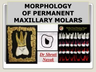
MORPHOLOGYOF PERMANENT MAXILLARY MOLARS Dr Shruti Nayak
There are three permanent maxillary molars. 1st, 2nd, and 3rd. FUNCTIONS: • Molar teeth have a major role in • Mastication of food • Support to the cheeks and also • Maintaining vertical dimension of face and fullness of cheek. INTRODUCTION
Maxillary molars are larger than other maxillary teeth • Crown is bucco-lingually larger in contrast to the corresponding teeth on the mandibular arch with are larger mesio-distally. General characteristics of Permanent Maxillary Molars
Crown is centered over the root and has 3 primary cusps and a fourth relatively smaller cusp that is disto-lingual cusp. • All maxillary molars have an oblique ridge extending from the most prominent mesio-lingual cusp to the disto-buccal cusp. • They have three roots, two buccal and one lingual. • All the roots converge to a root base called root trunk.
Arch traits • 3 roots: 2 B & 1 L • Crown: BL > MD • Cusps • 3 major cusps • ML, MB & DB • Lesser-sized DL cusp • Oblique ridge: ML to DB cusp • Buccal and lingual cusps are of unequal size • ML cusp is larger than DL MB DB ML DL Maxillary permanent molars
Class traits • 3 or more cusps • At least 2 buccal cusps • One or more lingual cusps • In general 2 or 3 roots Permanent Molars
Type trait (in general) • DL cusp reduces in size when going posteriorly & may be missing in 3rd molar • 1st molar is the largest & shows the least morphological variation • Cusp of Carabelli Maxillary permanent molars
Buccal aspect • MB & DB cusps are of equal height • MB is wider • Separated by B groove • Slopes of DB are steeper • Tip of the ML cusp between B cusps DB Maxillary permanent 1st molar MB
CROWN • Shape is trapezoidal with broader of the unequal part being occlusal and narrower the cervical outline. • Mesial out line- is nearly straight which becomes convex at mesial contact area located at the junction of occlusal and middle 1/3rd. • The out line continues to join to the mesial cusp slope of mesio-buccal cusp. M D BUCCAL ASPECT
DISTAL CONTACT AREA MESIAL CONTACT AREA • 1 • 2
Distal outlineis convex with contact area in the middle of middle 1/3rd • Occlusal outlineis represented by buccal cusps and cusp slopes. • From buccal aspect two cusps are seen; a mesio-buccal and disto-buccal cusps. • Mesio-buccal cusp is wider and slightly longer than the disto-buccal cusps. • Cervical outlineis irregular and shows slight curvature to the root. DB MB BUCCAL ASPECT
BUCCAL SURFACE • Buccal groove is seen separating two buccal cusps, extending to middle 1/3rd with a pit Roots • From the buccal aspect a distinct root trunk (undivided part of the root) is visible. • Root trunk bifurcate giving rise to two buccal roots a mesio-buccal and a disto-buccal root. • Both the roots are well separated, taper apically and often are curved distally. DB MB BUCCAL ASPECT
Crown – Crown of maxillary first molar is broader mesio-distally on the lingual side than on the buccal side • Two well developed cusps are visible from this aspect, the larger mesio-lingual cusp and smaller disto-lingual cusp. • Mesio-lingual cusp is the longest and largest cusp of this tooth and the cusp slopes meet at 90 degrees. • The disto-lingual cusp is smallest and is more rounded ML DL PALATAL ASPECT
PALATAL ASPECT 5TH D.L M.L
The lingual cusps are separated by a lingual developmental groove that extends from occlusal aspect to the lingual surface • Frequently a fifth cusp is found on the lingual surface of mesio-lingual cusp, located 2 mms cervical to the tip of the mesio-lingual cusp. • This cusp is separated from mesio-lingual cusp by a fifth cusp groove. • The cusp is named as ‘cusp of Carabelli’ after the person first described it. 5TH CUSP PALATAL ASPECT
The presence or absence of ‘cusp of Carabelli’ is a racial characteristic and when present may show variation in size & shape • Roots:Only one root is present on lingual side which is the longest of all 3 roots • The lingual root tapers to a blunt apex. • From the lingual aspect along with the lingual root both mesiobuccal and distobuccalroots are also visible. DL ML PALATAL ASPECT
Crown • Shape appears to the short and broad facio-lingually. • Buccal outlineis convex at cervical 1/3rd, followed by slight concavity and again convex as it progresses further to end at cusp tip. • Crestof buccal outline is usually located immediately below the cervical line. B L MESIAL ASPECT
Lingual outlineis some what similar to buccal outline, but crest of the lingual outline is often found at the middle 1/3rd • Cervical outlineis irregular and curved towards the crown • Mesial surfaceis generally convex. • A shallow concavity may be seen cervical to the contact area which may continue on to the root surface B L MESIAL ASPECT
crest crest L B
Occlusal outline– is represented by cusps and marginal ridge. • Two cusps are seen; a mesio-buccal cusp and a larger, longer mesio-lingual cusp. • Fifth cusp, the cusp of Carabelliis found on the lingual surface of mesio-lingual cusp. • Distinct mesial marginal ridge is present which is irregular and curved • Mesial marginal ridge is placed at an occlusal level than that of distal marginal ridge. MESIAL ASPECT
Root– Tow roots are visible from this aspect, mesio-buccal root and the lingual root. • The level of bifurcation on the mesial aspect is closer to(less than 4 mm) the cervical line. • The lingual root is 1.5 mm longer than MB root but narrower in a bucco-lingual direction. • The roots are well separated and the boundaries of the roots may extend beyond the crown. • This feature helps to differentiate this tooth from that of 2nd molar. Ling MB
Crown – Tooth shows a convergence distally making the buccal and lingual aspects visible • Mainly two cusps, disto-buccal and disto-lingual • Parts of other cusps including the ‘cusp of Carabelli’ can be seen. • Of the two cusps disto-buccal cusp is slightly larger than disto-lingual cusp. DL DB DISTAL ASPECT
The distal marginal ridge is shorter, more concave and cervically placed than mesial marginal ridge making a part of occlusal aspect visible from distal aspect. • Cervical time is less curved on distal aspect. • Distal surface is generally convex except for a shallow concavity at cervical region which may continue on to the root surface up to the level of bifurcation. DISTAL ASPECT
Roots:All the three roots are seen from this aspect. • The mesio-buccal root is seen because the disto-buccal root is shorter and narrow. • Lingual root seen • Level of bifurcation on the distal side is more apical than on mesial side. MB Ling DB DISTAL ASPECT
Occlusal outlineis rhomboidal / parallelogram in shape. • It has two acute angels and two obtuse • Acute angels are mesio-buccal and disto-lingual and obtuse angles are mesio-lingual and disto-buccal. O A OCCLUSAL ASPECT A O
Tooth is wider bucco-lingually (1 mm) than mesio-distally. • Crown shows a buccal and a distal convergence. • The lingual half of the tooth is wider mesio- distally than buccal half. • Similarly the mesial half of the tooth is bucco-lingually wider than distal half . OCCLUSAL ASPECT
Cusps - Four major cusps are seen; i.e. mesio-lingual cusp which is longest and largest followed by mesio-buccal, disto-buccal and disto-lingual cusp. • Of this four cusps mesio-lingual, mesio-buccal and disto-buccal forms the primary cusps of first molar. • A fifth cusp the ‘cusp of Carabelli’ is also seen lingual to mesio-lingual cusp which is located 2 mm cervical to the tip of the mesio-lingual cusp OCCLUSAL ASPECT
Square or rhomboidal • MB & DL angles are acute • ML & DB angles are obtuse • 1/3 of B surface & ½ of L are visible • B outline: D part is more L than M part OCCLUSAL ASPECT MB DB DL ML
BUCCAL • M • E • S • I • A • L • D • I • S • T • A • L • LINGUAL
BUCCAL • M • E • S • I • A • L • D • I • S • T • A • L • LINGUAL
Occlusal table • Cusps in order of decreasing size: ML, MB, DB & DL • M MR is longer & more prominent than D MR • Oblique ridge • Major Fossae • Central fossa • Central pit • Distal fossa • D pit • DL groove which continues as the L groove
CUSPS • M.B • D.B • 2 • 3 • 1 • 4 • M.L • D.L • 5
Ridges – Triangular ridges of all the four major cusps • The triangular ridge of the mesio-lingual cusp is divided into two parts by a groove named Stuart groove. • Distal extension of triangular ridge of the mesio-ligual cusp and of disto-buccal cusp meet and form a diagonal ridge called oblique ride. • Transverse ridge • Mesial and distal marginal ridges • Cusp ridges
RIDGES • TRIANGULAR RIDGES • M.B • D.B • OBLIQUE RIDGE • M.L • MARGINAL RIDGES • D.L TRIANSVERSE RIDGE
OCCLUSAL ASPECT • Fossae- There are four fossae on the occlusal aspect of a maxillary first molar, two major and other two are minor. • Major fossae :(a)Central fossa - largest fossa situated mesial to the oblique ridge, bounded by oblique ridge, transverse ridge and cusp ridges of buccal cusps. • (b) Distal fossais also a major fossa, relatively smaller than central fossa, and is located to distal to the oblique ridge. • It is linear in shape
CENTRAL FOSSA MTF DTF DISTAL
Minor fossae – • (a) Mesial triangular fossais a minor fossa, triangular in shape adjacent to mesial marginal ridge. • (b) Distal triangular fossasimilar to mesial triangular fossa, but smaller and is located adjacent to distal marginal ridge. • Pits – are observed at the deepest part of all fossae as pin point depression where the grooves converge OCCLUSAL ASPECT
Central groove– extends mesially from the central fossa, over the transverse ridge and ends in mesial triangular fossa. • Transverse groove of the oblique ridge. • This groove extends from the central fossa in a distal direction across the oblique ridge to the distal triangular fossa. GROOVES
Distal oblique groove– extends from the distal triangular fossa, along the distal aspect of oblique ridge in a lingual direction between the mesio-lingual and disto-lingual cusps. • Buccal groove – extends from the central fossa, in a buccal direction between the mesio-buccal and disto-buccal cusps and continues on to the buccal aspect of the tooth.
Lingual groove – This is seen as a continuation of the distal oblique groove and extends on to the lingual surface of the tooth between mesio-lingual and disto-lingual cusps. • Fifth cusp groove – separates the fifth cusp from the mesio-lingual cusp. • Stuart groove – This is a small groove which extends from central groove to separates the two portions of triangular ridge of mesio-lingual cusp.
Transverse groove of the oblique ridge • Buccal groove • Central groove Grooves Stuart groove • Fifth cusp groove • Distal oblique groove • Lingual groove
Pulp • MD section • 2 horns, MB is higher • Pulp chamber, roof & floor • Canals, narrow • Canal orifice • BL section • Pulp chamber is wider • 2 horns of equal height • X-section • 3 canals Maxillary permanent 1st molar
Maxillary second molars are situated distal to the first molars. • They assist first molars in function. • These teeth may show considerable variation in morphology. Maxillary second molar
Buccal aspect (type traits) • Smaller crown size • Less prominent DB cusp & narrower MD • Distally inclined B roots • Lingual aspect • DL cusp is smaller in width & height • L root is narrower MD & slightly D inclined • No cusp of Carabelli Maxillary permanent 2nd molar
L MB DB DISTAL MESIAL DB MB
Crown • Crown is shorter and less widerthan first molars and is tipped distallyon the root trunk. • Mesial outline is slightly convex with contact area located at the junction of occlusal and middle 1/3rd. • Distal outline is shorter than the mesial outline. • Distal contact area is located at the middle of middle 1/3rd. Buccal aspect