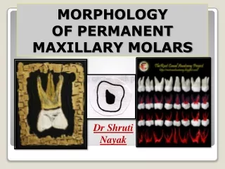
Permanent Maxillary Molars
520 likes | 696 Vues
Permanent Maxillary Molars. Maxillary First Molar. General Characteristics. Arch position: 6th tooth from midline Universal #3 and #14 Mesially, contacts primary 2nd molar and later 2nd premolar Largest tooth in maxillary arch Largest crown in mouth

Permanent Maxillary Molars
E N D
Presentation Transcript
General Characteristics • Arch position: 6th tooth from midline • Universal #3 and #14 • Mesially, contacts primary 2nd molar and later 2nd premolar • Largest tooth in maxillary arch • Largest crown in mouth • Least variable in anatomic form among max molars
General form: • Three roots: L, MB, DB • Slightly wider F-L than M-D • Trapezoidal geometric form F/L and proximal views • Occlusal view: rhomboidal geometric form (described as 5-sided) mesial facial
Development Timeline: • Initial calcification: at birth* • Enamel completed: 3 - 4 years • Eruption: 6 - 7 years • Root completed: 9 - 10 years
Facial view, mesial outline: • Mesial HOC at junction of occlusal and middle thirds • Outline relatively flat cervical to mesial HOC • M-D > O-C dimension
Facial view, distal outline: • Distal HOC at middle third • Entire distal outline convex • Disto-occlusal angle more rounded than mesio-occlusal angle • Distal surface visible from facial view (rhomboidal effect)
Facial view, cervical line: • Oftentimes a V-shape dip CE line pointing towards buccal bifurcation
Facial view, occlusal portion: • MB and DB cusps separated by buccal groove • MB cusp wider, DB cusp tip sharper* (sharpest of 4 cusps) • ML cusp visible between MB and DB cusps
More facial… • Buccal groove usually ends midway O-C in a buccal pit • Bucco-gingival ridge runs horizontally, more prominent towards mesial • Facial HOC at cervical third
Lingual View • DLG separates larger ML cusp from smaller DL cusp • DLG oftentimes terminates in lingual pit • Lingual groove midway M-D • M-D width as wide or wider lingually than facially*
Lingual view… Georg Carabelli court dentist to Austrian emperor (1842 A.D.) • Cusp of Carabelli* • Lingual surface of ML cusp • Varies in size: non-existent to prominent • Lingual HOC at middle third • Lingual root depression*
Mesial view, facial outline: • Buccal HOC at cervical third (bucco-gingival ridge) • Facial surface fairly flat from HOC to cusp tip
Mesial view, lingual outline: • Lingual HOC at middle third • Overall convex curvature • Palatal root apex is lingual to the crown
Mesial view, occlusal outline: • Only MB and ML cusps visible • Mesial marginal groove usually present, midway of marginal ridge • Mesial contact area situated buccal to mesial groove, at junction of occlusal and middle thirds
Distal View • More of occlusal surfaces visible • All four cusp tips visible • Distal marginal groove usually present, midway of marginal ridge • Pronounced distal cervical crown/root concavity*
Distal view… • More of facial surface visible, but less of lingual surface (rhomboidal form) • Distal contact area located midway F-L, more cervical than mesial contact (middle third) Mesial view
Occlusal View • Rhomboidal geometric form • Acute MB and DL corners • Obtuse DB and ML corners • 5-sided (pentagonal) • Largest F-L dimension of any tooth*
Occlusal view… • F-L and M-D dimensions more nearly similar* • M-D width as wide or wider lingually than facially* • Four major cusps: ML, MB, DB, DL (largest to smallest) • Cusp triangle: MB, DB, ML (trigon)
Occlusal view… • Transverse ridge • Triangular ridges of MB and ML cusps • Oblique ridge • Triangular ridges of DB and ML cusps (distal cusp ridge?) • About same height as the marginal ridges* • Mesial 2/3 looks like max premolar
Occlusal view… • Three pits: mesial, central, distal • Three primary developmental grooves: central, buccal, distolingual • Four fossae: mesial, central, distal, distolingual
Root Form • Root trunk with trifurcation: • Lingual root: • Largest, longest • Wider M-D than F-L* • MB root • 2nd largest • Apex in line with MB cusp tip* • DB root • Smallest of three
Root form... • DB root area may have pronounced cervical concavity* • MB and DB root forms look like “plier handles” • MB root could have two pulp canals (70% possibility) • Root depression, lingual surface of lingual root
How To Tell Right From Left: • Distolingual groove • Cusp of Carabelli on ML cusp • Broader MB root than DB root • MB prominence of facial surface
General Characteristics • Arch position: 7th from midline • Universal #2 and #15 • Wider F-L than M-D • Similar to 1st, except smaller • Smaller DL cusp
Development Timeline: • Initial calcification: 2½ - 3 years • Enamel completed: 7 - 8 years • Eruption: 12 - 13 years • Root completed: 14 - 16 years
Facial View • Narrower M-D than 1st • M-D > O-C dimension • MB cusp larger than DB • Buccal groove more distally located than 1st • MB root tip in line with buccal groove*
Lingual View • Smaller DL cusp than 1st • Lingual groove more distally located • No Cusp of Carabelli
Mesial and Distal Views • Similar to 1st except shorter O-C • Facial HOC at cervical third • Lingual HOC at middle third • Mesial HOC at junction of occlusal-middle • Distal HOC slightly more cervical
Occlusal View • Narrower M-D than F-L • Tapers lingually • Two major crown forms: - Rhomboidal - Heart-shaped (diminished DL cusp)
Root Form • MB and DB roots closer than 1st • MB apex in line with buccal groove* • No lingual root depression
How To Tell First From Second: • Occlusal view of first less rhomboidal than second • Cusp of Carabelli only on first • M-D and F-L dimensions more similar with first, narrower M-D with second*
First from second... • MB root apex in line with MB cusp tip with first, in line with buccal groove with second*
General Characteristics • Arch position: 8th from midline • Universal #1 and #16 • No distal contact area • Only maxillary tooth that has single opposing tooth* • Smallest of all molars - shortest O-C dimension of any tooth*
Development Timeline: • Initial calcification: 7 - 9 years • Enamel completed: 12 - 16 years • Eruption: 17 - 21 years • Root completed: 18 - 25 years
Crown Form • Most variable of maxillary posteriors • Heart-shape most common • Diminished or absent DL cusp • 3-cusp form (L, MB, DB cusps) • Still wider F-L than M-D • Occlusal table more constricted • No distal contact wear facet
Root Form • Three roots, partially or fully fused • Roots much shorter than 1st and 2nd, crown:root ratio closer to 1:1 • Roots shorter than any other tooth (other than mandibular 3rds)
How To Distinguish a Third: • Shorter root: Crown to root ratio nearly 1:1* • Roots usually fused • More supplemental occlusal grooves* • Heart-shape occlusal outline, narrower occlusal table
How To Tell Right From Left: • MB root larger than DB root • MB prominence of facial surface • DLG, if present • Contact area wear facet only on mesial