Understanding the Human Heart: Structure, Function, and Blood Circulation
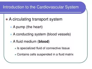
This comprehensive guide explores the anatomy and function of the human heart. It covers the heart’s four chambers, associated blood vessels, and valves, as well as the pathway of blood flow. The guide explains coronary artery function, myogenic contraction, pacemaker activity, and nerve control of the heartbeat. It outlines the structure-function relationship of arteries, capillaries, and veins, and details components of blood, including plasma and blood cells. Additionally, it describes the pulmonary and systemic circulation processes, emphasizing the heart's dual pumping system.

Understanding the Human Heart: Structure, Function, and Blood Circulation
E N D
Presentation Transcript
Assessment Statement • 6.2.1 Draw and label a diagram of the heart showing the four chambers, associated blood vessels, valves, and the route of blood through the heart • 6.2.2 State that the coronary arteries supply heart muscle with oxygen and nutrients • 6.2.3 Explain the action of the heart in terms of collecting blood, pumping blood, and opening and closing of valves • 6.2.4 Outline the control of the heartbeat in terms of myogenic muscle contraction, the role of the pacemaker, nerves, the medulla of the brain and adrenaline • 6.2.5 Explain the relationship between the structure and function of arteries, capillaries, and veins • 6.2.6 State that blood is composed of plasma, erythrocyctes, leucocytes (phagocytes and lymphocytes) and platelets • 6.2.7 State that the following are transported by the blood: nutrients, oxygen, carbon dioxide, hormones, antibodies, urea and heat
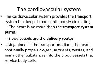
The Human Heart • The human heart is designed as a pair of side by side pumps • Each side of the heart has a collection chamber for blood that is moving slowly in from the veins. • These are called atria (thin walled) • Each side also has a thick walled muscular pump which builds up enough pressure to send the blood out from the heart with a force (blood pressure) • These are called ventricle
The Human Heart • The blood that is pumped out from the heart typically makes a circuit through the following range of blood vessels • A large artery • Smaller artery • An arteriole • A capillary bed • A venule • Larger veins • A large vein which takes blood back to the heart to be pumped out once again
The Human Heart • The two sides of the heart allow for there being two routes for blood to flow along • The right side of the heart sends blood along a route that is called your pulmonary circulation • One this route, the capillary bed is in one of your lungs, and blood picks up oxygen and releases carbon dioxide
The Human Heart • The left side of the heart sends blood along a route that is called your systemic circulation • The artery that emerges from your heart for this route is your aorta • Branches of the aorta carry blood to almost every organ and cell type in your body • On this route, the capillary bed is in one of your organs or tissues, and blood picks up carbon dioxide and releases oxygen
Pulmonary Circulation • Imagine a red blood cell • The blood cell is first found in a large vein that is bringing blood to the right atrium • Because it is already been to the body tissues it needs more oxygen
Pulmonary Circulation • A volume of blood collects within the right atrium and begins moving down into the right ventricle through a open valve • This is the right atrioventricular valve
Pulmonary Circulation • The right atrium contracts in order to force any remaining blood into the right ventricle • This contraction initiates several events including: • Closure of the atrioventricular valve to prevent backflow to the right atrium (it is the closing of valves that produces the characteristic ‘lub dub’ sounds heard through a stethoscope
Pulmonary Circulation • Dramatic increase in blood pressure inside the right ventricle which opens the right semilunar valve and allows blood to enter the pulmonary artery • Due to the increase in pressure, blood leaves the heart through the pulmonary artery
Pulmonary Circulation • Our RBC is now in an artery leading to one of the two lungs • As it approaches and then enters a lung, our RBC will be moving along smaller and smaller arteries • Any one arteriole leads to a capillary bed • Capillaries are blood vessels that have a very small diamter and are typically only a single cell thick • This is why exchanges only while blood is in capillaries • Then the RBC will be on its way back to the heart. It pass into larger and larger veins until the largest of those veins takes our RBC directly into the left atrium
Systemic Circulation • When our RBC enters the left atrium, a set of events occurs that is similar to when it entered the right atrium • In fact, the right and left sides of the heart are acting in unison—both atria contract at the same time and both ventricles contract at the same time • Blood, now including our RBC, accumulates in the left atrium and then enters the open, left atrioventricular valve • Our RBC passes into the left ventricle as this chamber fills with blood
Systemic Circulation • When the left ventricle contracts, this initiates the following events: • Closure of the atrioventricular valve to prevent backflow into the left atrium • Dramatic increase in blood pressure inside the left ventricle when opens the left semilunar valve and allows blood to enter the aorta • Due to the increase in pressure, blood leaves the heart through the aorta
Systemic Circulation • Our chosen RBC now finds itself in the largest artery in the human body • The aorta has many branches which lead to all tissues in the body • One of the first branches from the aorta allows blood to enter the coronary arteries. The coronary arteries branch out into the heart muscle itself and supply the heart with oxygen and nutrients • Now our RBC ultimately finds itself in a capillary bed somewhere in the body. • This is where oxygen is given off and carbon dioxide may be taken in by the blood
Circulation • Each complete circuit round the body includes both the systemic route or circuit and the pulmonary route or circuit. • Each complete circuit typically takes not longer than a minute or two
Control of Heart Rate • The majority of the tissue making up the heart is muscle (cardiac muscle) • Cardiac muscle spontaneously contracts and relaxes without nervous system control • This is known as myogenic muscle contraction-The myogenic activity of the heart needs to be controlled in order to keep the timing of the contractions unified and useful
Control of Heart Rate • The right atrium contains a mass of tissue within its walls known as the sinoatrial node (SA node) • This mass of tissue acts as the pacemaker of the heart • It sends out an ‘electrical’ signal to initiate the contraction of both atria • Normal resting heart rate, the signal from the SA node is sent out every 0.8 seconds
Control of Heart Rate • Also within the right atrium, is another mass of tissue known as the atrioventricular node (AV node) • The AV node receives the signal from the SA node, waits approximately 0.1 seconds and then sends out another ‘electrical’ signal • This second signal goes to the much more muscular ventricles and results in their contraction • This explains why both atria and then, later, both ventricle contract together
Exercise • During times of increased body activity, the heart rate needs to increase above the resting heart rate. • This is because there is an increased demand for oxygen for cell respiration during periods of heavy exercise or body activity • There is also a need to get ride of the increased levels of carbon dioxide that accumulate in the bloodstream • This increase level of CO2, an area of the brainstem called the medulla chemically senses the increase in CO2 • The medulla then sends a signal through a cranial nerve to increase heart rate to an appropriate level
Exercise • This signal is sent to the SA node, it does not change the mechanism of how the heart beats, just the timing • After exercise, the level carbon dioxide in the bloodstream begins to decrease and another signal is sent from the medulla. • This time the signal is carried by a different craniel nerve called the vagus nerve • Ultimately, electrical signals from the vagus nerve result in the SA node once again taking over the timing of the heart rate
Chemicals • The heart rate can also be influenced by chemicals • Most common is adrenaline • During periods of high stress or excitement, your adrenal glands secrete adrenaline into the bloodstream • Among other effects, adrenaline causes the SA node to ‘fire’ more frequently than it does at the resting heart rate and thus heart rate increases
Arteries, capillaries and veins • Arteries are blood vessels taking blood away from the heart that has not yet reached a capillary • Arteries have a relatively thick smooth muscle layer that is used by your autonomic nervous system to change the inside diameter of the blood vessels. This helps in regulating blood pressure
Arteries, capillaries and veins • Veins are blood vessels that collect blood from capillaries and return to the heart • Receive blood at relatively low pressure from capillary beds. Because this blood has lost a great deal of blood pressure, the blood flow through veins is slower than through arteries • To account for this, veins have thin walls and a larger internal diameter • Veins also have many internal passive valves that help keep the slow moving blood consistently moving towards the heart • Capillary bed is a network of capillaries that typically all drain into a single venue • When blood enters a capillary bed much of the pressure is lost. Blood cells make their way through capillaries one cell at a time • Chemical exchanges occur
Arteries, capillaries and veins • Capillary bed is a network of capillaries that typically all drain into a single venue • When blood enters a capillary bed much of the pressure is lost. Blood cells make their way through capillaries one cell at a time • Chemical exchanges occur