Visual System
Visual System. Overview of Pathways – Functional Anatomy. Another sensory system, like touch and pain. Therefore, organized in similar way: thalamus sensory system (retina) ------- 1° visual cortex.

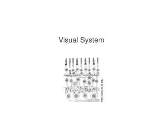
Visual System
E N D
Presentation Transcript
Overview of Pathways – Functional Anatomy • Another sensory system, like touch and pain. • Therefore, organized in similar way: thalamus sensory system (retina) ------- 1° visual cortex. As in other sensory systems, projections to thalamus and visual cortex is very topographically organized.
The right visual field maps on the left visual cortex and vice versa Neural pathway Figure 10.36
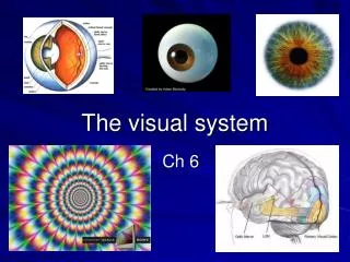
Retina 1. Retina regions 2. Retina layers and cell types. B. Optic nerve and optic chiasm. C. Superior Colliculus. D. Lateral geniculate nucleus (of the thalamus). E. Optic radiations and projections to the 1° visual cortex. 1. magnocellular/parvocellular projections. 2. Column Organization of the 1° visual cortex. a. ocular dominance columns. b. orientation columns. c. color blobs. F. Inputs to higher-order visual cortical areas. Neural pathway – Regional Anatomy of the Visual System
Photoreceptor cells Inter-neurons Ganglion cells Optic nerve Optic chiasm Optic tract 2 Primary Visual Paths (See Fig. 7-1) LGN Brachium of superior colliculus Optic radiations Superior colliculus 1° visual Cortex (calcarine fissure)
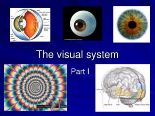
Pathway for Visual Perception: (Fig. 7-1A) • Inputs to the 1° visual cortex • serve basic visual perception. • After this initial input, pathways • may then ascend to 2° and higher- • order cortical areas in • surrounding parts of the occipital • lobe. • These other parts serve other aspects • of vision, such as colour vision • and visual motion. • A decussating path (through corpus • callosum) terminates on contralateral 1° • cortex (unify images from 2 eyes). • A descending path goes to • oculomotor centres in midbrain, in order • to focus visual image on the retina.
Pathway for Control of Eye Movement (Fig. 7-1B): For control of eye movements: Pathway to midbrain. Superior colliculus: inputs in voluntary control of saccades (REMs to salient stimuli) and in coordinating movements of head in concert with this. Pretectal nuclei participate in pupillary reflexes
Regional Anatomy of the Visual System • Retina and optic nerve are derived from out-pocketings of the diencephalon and are, therefore, part of the CNS, and not the PNS (in case you were wondering). • As seen in Fig. 7-1B, temporal half of each eye projects ipsilaterally, where as the nasal half projects contralaterally. • Optic disk – where axon exits and blood vessels pass – a “blind spot”. • Macula – area of highest visual acuity. • Fovea – centre of the macula. • Retinal layers and cell types (see preceding and following slide)
Photoreceptor cells Inter-neurons Ganglion cells Optic nerve Optic chiasm Optic tract 2 Primary Visual Paths (See Fig. 7-1) LGN Brachium of superior colliculus Optic radiations Superior colliculus 1° visual Cortex (calcarine fissure)
3 cell layers, 2 synaptic layers: Outer layer: photoreceptors- rods (night vision) and cones (for daylight and colour, densest just around the fovea) Middle layer: bipolar neurons: horizontal (more superficial) and amacrine (deeper). Both perform lateral interactions, which enhance visual contrast. Inner layer: ganglion cells. Note that light goes through to outermost layer hits photoreceptor cells. Regional Anatomy of the Visual System: Retinal structure
Cone bipolar cells: Input from small # Of cones for high visual Acuity. Rod bipolar cells: Input from several rods for convergence and increased sensitivity at low illumination. At outermost extreme: Pigmented epithelium – Phagocytic role for removing old photoreceptors. This function is absent in retinosa pigmentosum, which can detached retina
Neural processing • The bipolar neurons and ganglion cells process the signal • In the fovea where the acuity is the highest: 1 cone 1 bipolar cell 1 ganglion cell • At the periphery: many rods 1 bipolar cell … acuity is much decreased • Other cells in the retina participate in signal processing
Optic Nerve and Optic Chiasm • Ganglion cell axons (which are clear and unmyelinated, while running along the inner surface of the retina) gather together and exit at the optic disk, where they become myelinated and form the optic nerve (actually, the 2nd cranial nerve, but since the CNS, officially a tract). • Optic nerves from both eyes converge at optic chiasm: partial cross-over. • Images in the nasal hemiretina from both sides cross over (temporal stay ipsilateral). • This allows for complete cross-over of each visual field (see Fig. 7-3C).
The right visual field maps on the left visual cortex and vice versa Neural pathway Figure 10.36
Superior colliculus • As noted above, some optic tract axons do not go to the LGN of the thalamus, but instead, travel in the brachium of the superior colliculus. • Laminated - 1st layer receives direct input from the retina (brachium). Other layers receive input from 1st or from somatic sensory (AL system) and auditory systems. - sensory info from the different modalities is lined up in the different layers output to eye and neck muscles so that one can properly orient to the stimulus.
Superior colliculus
Lateral Geniculate Nucleus A nucleus in the thalamus, which projects to the 1° visual cortex and serves visual perception. A recurrent theme: this nucleus is also laminated (6 layers) with alternating input from ipslateral and contralateral retina. There is also a division between 2 important input systems: Magnocellular – input from M ganglion cells with wide dendritic arbours (integrates visual input info from wide area for motion vision). Parvocellular – input from P cells (ganglionic cells with small dendritic arbours) – for discriminative aspects of visual form and colour. These systems project to distinct parts of the 1° visual cortex.
Optic Radiations and Projections to the Primary Visual Cortex • Meyer’s Loop: course anterior for a short distance in order to move over the lateral ventricles. • 1° cortex, in columnar fissure, is also Brodman’s Area 17. • Densest projection to the 1° visual cortex (like other sensory) is to Layer 4 (Stripe of Gennari).
Inputs to primary visual cortex Parvocellular Magnocellular Note: both have distance Projections and destinations. These, in turn, project to higher- order areas for visual processing.
Columnar Organization Neurons lined up with similar properties across the different layers. e.g., somatic sensory cortex: columns from same location in body and same modality. In visual system, 3 types of aggregates (2 transverse the whole thickness of cortex and, therefore, are considered columns: • Ocular dominance columns. • Orientation columns. • Aggregates of colour-sensitive neurons (“colour blobs”).
Orientation columns: inputs with similar spatial orientation
“Colour blobs”: aggregates of colour-sensitive neurons (layers 2 and 3 only)
Inputs to Higher-Order Visual CorticalAreas • Higher-order areas surround area 17 (areas 18 and 19), and can be distinguished as they lack a Stripe of Gennari. • Motion pathway – Magnocellular system 1° cortex higher-order areas for visual form in motion (including medial temporal projection). • Colour pathway – parvocellular system 1° cortical projections in 4Cβ colour blobs higher-order areas. • Form visual pathway – parvocellular system 4Cβ region between blobs higher-order cortex and inferior temporal lobe. Two “streams” of projections outside the visual system: - ventral: features “what” - dorsal: spatial information: “where”
Visual Field Deficits after Lesions at Various Levels of the Visual System • Optic n.: transection = monocular blindness. • Optic chiasm: (e.g., pituitary tumour). Transection bitemporal visual field deficit. • Optic tract or LGN: transection contralateral visual field deficits (homonymous hemianopsis). • Optic radiations: transection of Meyer’s Loop only (temporal region) contralateral upper quadrant (‘quadranotopia’). Complete transection homonymous hemianopsia. • 1° Visual Cortex: most common = infarction homonymous hemianopsia with macular sparing (2° greater cortical representation).
What will happen if the left optic nerve is severed? What will happen if a person has a tumor in the pituitary gland (just below the optic chiasmata) and the inner fibers are destroyed? What will happen if a person suffers a brain tumor on the right side of the brain around the lateral geniculate body? Neural pathway – Regional Anatomy of the Visual System