Visual System
Visual System. http://www.cis.rit.edu. Much of the text material is from, “Principles of Anatomy and Physiology (12th edition)” by Gerald J. Tortora and Bryan Derrickson (2009). I don’t claim authorship. Other sources are noted when they are used.

Visual System
E N D
Presentation Transcript
Visual System http://www.cis.rit.edu
Much of the text material is from, “Principles of Anatomy and Physiology (12th edition)” by Gerald J. Tortora and Bryan Derrickson (2009). I don’t claim authorship. Other sources are noted when they are used. Mapping of the lecture slides to the 13th edition is provided in the supplement.
Outline • Introduction • Nature of light • Basic eye structure • Image projection—retina • Image processing and interpretation
Importance of Vision • Vision—which is very important for human survival—uses substan-tial neural resources. • More than 50 percent of the sensory receptors of the human body are located in the eyes. • Large areas of the cerebral cortex process and interpret visual infor-mation. • As many as 30 cortical areas are involved in the processes of visual processing and interpretation. Neural = of or relating to a nerve or the nervous system. Chapter 17, page 604
Electromagnetic Radiation • Electromagnetic radiation is energy manifested in the form of particles (photons) and waves that radiate from the sun and other light sources. • Its vast range of wavelengths is called the electromagnetic spectrum. • The distance between two consecutive wave peaks is its wavelength. • Wavelength is used to calculate frequency, that has a unit of measure-ment known as Hertz. Hertz (Hz) = number of complete cycles per second. Figure 17.4 Chapter 17, page 605
Electromagnetic Spectrum (continued) Increasing energy Wavelength 10-5 nm 10-3 nm 1 nm 103 nm 106 nm 1 m 103 m Electromagnetic spectrum Gamma rays Ultra- violet X-rays Infrared Microwaves Radio waves 380 nm 750 nm Visible light VioletIndigoBlueGreenYellowOrangeRed The light we see is a very small slice of the electromagnetic spectrum. Sunlight is 45 percent visible light, 46 percent infrared radiation, and 9 percent ultraviolet radiation.
Wavelength • Wavelengths of electromagnetic radiation range from very short to very long: • Gamma rays are shorter than a nanometer (nm), or billionth of a meter. • Radio waves exceed one meter. http://www.nasa.gov Figure 17.4 Chapter 17, page 605
Visible Light • Visible light is the part of the electromagnetic spectrum that ranges from about 400 to 700 nm. • Color sensation depends on the wavelength of light and its processing by the visual system. • For example, the color violet is associated with a wavelength of about 400 nm, while red is associated with a wavelength of about 700 nm. Figure 17.4 Chapter 17, page 605
A Mnemonic Device • From longest to shortest wavelengths of visible light, use the mnemonic, ROY G BIV. • R = red • O= orange • Y= yellow • G = green • B = blue • I = indigo • V = violet http://www.missouriskies.org
Other Animals • Animals may have somewhat different sensitivities to the electromag-netic spectrum. • Pit vipers (such as rattlesnakes) are sensitive to infrared radiation to track prey at night. • Honeybees are sensitive to ultraviolet radiation to navigate to a nectar source—see the book, “The Dancing Bees” by the ethologist, Karl von Frisch. • We, as humans, are not able to detect infrared and ultraviolet radiation without special instruments.
Absoption and Reflection • Objects absorb some wavelengths and reflect others—an object will appear as the colors associated with the wavelengths that are reflected from it. • A new pair of blue jeans absorbs all wavelengths of the visible spec-trum except those associated with blue, which are reflected. • An object appears white because it reflects all lengths of visible light. • An object appears black because it absorbs all wavelengths of visible light. http://featuresblogs.chicagotribune.com Chapter 17, page 605
Basic Eye Structure http://www.wellhappypeaceful.com
Extrinsic Eye Muscles • The extrinsic eye muscles extend from the walls of the bony orbit to the sclera (white) of the eye. • The muscles are controlled by the oculomotor (III), trochlear (IV), and abducens (VI) cranial nerves. Figure 17.6 Chapter 17, page 606
Eye Movements • The six extrinsic eye muscles are capable of moving the eye in almost any direction. • They can move the eyeball laterally (to the side), medially (away from the side), superiorly (up), and inferiorly (down). • Another set, known as the oblique muscles, enable rotational stability of the eye. Figure 17.6 Chapter 17, page 606
Motor Units • The motor units of the extrinsic muscle fibers of the eyes are very small. • Some motor neurons serve only two or three extrinsic muscle fibers, fewer than in any other part of the body except the larynx, or voice box. • The small motor units permit smooth, rapid, and precise movements. • Neural circuits in the brainstem, including in the superior colliculi and the cerebellum, coordinate and synchronize eye movements, such as the scanning of a visual scene. Chapter 17, page 606
Eyeball • The adult eyeball measures about 2.5 cm (one inch) in diameter. • About one-sixth of the eyeball’s total surface area is exposed, and the rest is protected by the bony orbit. • The wall of the eye consists of three layers—fibrous tunic, vascular tunic, and retina. Figure 17.7 Chapter 17, page 606
Eyeball (continued) http://www.blundelloptometry.com
Fibrous Tunic • The fibrous tunic, the superficial layer, consists of the cornea and the sclera. • The cornea is a transparent tissue that covers the colored iris—it helps focus light on the retina. • The sclera—the white of the eye—is a dense layer of connective tissue formed mostly from collagen fibers and fibroblasts. Superficial = outer. Figure 17.7 Chapter 17, page 607
Fibrous Tunic (continued) • The sclera covers the entire eyeball except the cornea. • It gives shape to the eyeball, makes it more rigid, protects its inner components, and serves as attachment points for the extrinsic eye muscles. Figure 17.7 Chapter 17, page 607
Vascular Tunic • The vascular tunic—the middle layer—consists of the choroid, ciliary body, and iris. • Blood vessels of the choroid provide nutrients to the posterior surface of the retina. • The choroid contains melanocytes that produce the pigment melanin, giving it a dark brown color. • Melanin absorbs stray light rays to: 1) prevent reflection and scattering of light within the eye, and 2) help assure that the image projected onto the retina is sharp and clear. Figure 17.7 Chapter 17, page 608
Vascular Tunic—Ciliary Body • The anterior portion of the vascular tunic becomes the ciliary body. • The protrusions and folds on the internal surface of the ciliary body, known as ciliary processes, secrete a fluid known as aqueous humor. • A circular band of smooth muscle fibers, the ciliary muscle, changes the shape of the lens for both near- and far-vision. Chapter 17, page 609
Vascular Tunic—Iris • The colored portion of the eye, the iris, is shaped like a flattened donut. • It is suspended between the cornea and the lens, and is attached at its outer margin to the ciliary processes. • The iris contains melanocytes and circular and radial smooth muscle fibers. http://www.army-technology.com Chapter 17, page 609
Vascular Tunic—Iris (continued) • The amount of melanin in the melanocytes in the iris determines eye color: • Brown-to-black color with a large concentration of melanin. • Green when the concentration is moderate. • Blue when the concentration is very low. • The iris regulates the amount of light entering the eye through the pupil. Chapter 17, page 609
Pupil • The pupil appears black due to the heavily-pigmented choroid and the retina. • If bright light is directed into the eye, such as with an ophthalmoscope, the reflected light is red due to the blood vessels on the surface of the retina. • A person’s eyes will appear red in a photograph if the camera flash is directed into the pupil. Ophthalmoscope = an instrument for examining the interior structures of the eye, especially the retina. Chapter 17, page 609
Pupil (continued) • Autonomic reflexes regulate the diameter of the pupil in response to light levels, arousal, and emotional states. • The parasympathetic fibers of the oculomotor (III) cranial nerve stim-ulate the circular muscles to contract, producing constriction. • Sympathetic fibers stimulate the radial muscles to contract, producing dilation. Figure 17.8 Chapter 17, page 609
Retina • The retina, the inner layer of the eye, lines the posterior three-quarters of the eyeball. • It serves as the first stages of visual processing by the nervous system. • An ophthalmoscope can be used to view the retina, which is the only place where blood vessels can be viewed directly in the body to detect pathological changes. Figure 17.9 Chapter 17, page 609
Retina—Visual Appearance Ophthalmoscope http://jbooy.files.wordpress.com Retina http://medrninja.files.wordpress.com
Retina (continued) • Retinal changes can result from hypertension, diabetes mellitus, cata-racts, and age-related macular degeneration. Cataract = an eye disease that involves the clouding, or opacification, of the lens. Macular degeneration = chronic eye disease marked by deterioration of the tissue responsible for central vision. Chapter 17, page 609
Retina—Prominent Features • Several features of the retina are visible through an ophthalmoscope: • Optic disc, where the optic (II) cranial nerve exits the eye • Central retinal artery and vein • Macula lutea and fovea • Branches of the central retinal artery fan-out to nourish the anterior surface of the retina. • The central vein drains blood from the retina through the optic disc. Figure 17.9 Chapter 17, page 609
Pigmented Layer • The retina consists of a pigmented layer, and a neural layer of special-ized nerve cells. • The pigmented layer, located between the choroid and neural layer, has epithelial cells that contain melanin. • The melanin concentration in the pigmented layer, as in the choroid, ab-sorbs stray light. Chapter 17, page 610 Figure 17.10
Neural Layer • The neural or sensory layer of the retina is a multi-layered outgrowth of the brain. • The photoreceptors and neurons in the neural layer begin processing visual information before it is transmitted via the optic nerve to the thalamus and superior colliculi. • Retinal neurons are in three distinct sub-layers, separated by synaptic connections. Figure 17.10 Chapter 17, page 610
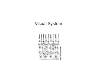
Cross Section of the Retina Rod and cones Outer synaptic sub-layer Horizontal cell Bipolar cells Amacrine cells Inner synaptic sub-layer Ganglion cells Adapted from http://www.skidmore.edu Direction of light
Neural Layer (continued) • The neural layer has sub-layers of photoreceptors, bipolar cells, and ganglion cells. • Light must pass through the bipolar and ganglion sub-layers before it reaches the photoreceptors. Figure 17.10 Chapter 17, page 610
Neural Layer (continued) • Besides bipolar cells, the bipolar sub-layer has horizontal cells and amacrine cells. • These cells have lateral synaptic connections that modify the neural signals transmitted from the photoreceptors to the bipolar cells, and then to the ganglion cells. • This arrangement, as will be discussed, enables visual processing to begin in the retina. Figure 17.10 Chapter 17, page 610
Photoreceptors—Rods • Photoreceptors—the rods and cones—begin the process in which visible light is converted to action potentials for transmission to the brain. • Each human eye has about 120 million rods and 6 million cones. • Rods enable vision in dim light—they provide sensations of shades of gray, but not color. Figure 17.10 Chapter 17, page 610
Photoreceptors—Cones • The cones, which enable color vision, are active at higher levels of illumination. • The three types of cones are sensitive to either blue, green, or red light. • Color vision results from the stimulation of combinations of the three types of cones to produce the full palette of colors that we experience. • All of the colors that we perceive result from stimulation of red, blue, and green receptors. Chapter 17, page 610
Rods and Cones www.eyedesignbook.com Electron micrograph
Information Flow • From the photoreceptors, visual information flows through an outer synaptic sub-layer to the bipolar cells. • The information then flows through an inner synaptic sub-layer to the ganglion cells. • The axons of the ganglion cells enter the optic disc and exit the eye as the optic (II) cranial nerve. Figure 17.10 Chapter 17, page 610
Blind Spot • The optic disc contains no rods or cones. • We rarely notice the blind spot since the brain fills in the information gap. • The blind spot is located slightly off-center from the visual axis so that vision is not severely hindered when a person looks directly at an object. • The textbook contains a simple demonstration for finding your blind spot in each eye. Figure 17.10 Chapter 17, page 610
Macula Lutea and Fovea Centralis • The macula lutea is at the exact center of the retina, along the visual axis of the eye. • The fovea centralis—a small depression in the center of the macula lutea—contains cones but no rods. • The sub-layers of bipolar and ganglion cells, which could scatter the incoming light, are displaced to the side of the fovea centralis. • The fovea centralis is the area of highest visual acuity (resolution) due to its high density of cones. Figure 17.9 Chapter 17, page 610
Fovea Centralis—Cross Section http://dspace.udel.edu Electron micrograph
Fovea Centralis (continued) • A primary reason for moving one’s head and eyes when looking at an object is to position the image on the fovea centralis to maximize visual acuity. • Rods are absent from the fovea centralis, but are abundant toward the periphery of the retina. • Because rods are more sensitive to light than cones, a faint object (such as a dim star) is better viewed if a person shifts her or his gaze slightly to the side, away from the fovea centralis. Chapter 17, page 610
Lens • The lens is located in the cavity of the eye behind the pupil and iris— its role is to help focus visual images on the retina in conjunction with the cornea. • Crystallins (proteins) within the lens cells are arranged like layers of an onion to help form the refractive media. • The lens lacks blood vessels and is normally transparent. • The lens is enclosed in a clear connective tissue capsule, and is held in place by zonular fibers attached to the ciliary processes. Refractive media = includes the lens and vitreous humor. Figure 17.7 Chapter 17, page 610
Interior of the Eye • The lens divides the interior of the eye into the anterior and vitreous cavities. • The anterior cavity consists of anterior and posterior chambers. • The anterior chamber lies between the cornea and iris. • The posterior chamber is located behind the iris and in front of the lens and zonular fibers. Figure 17.11 Chapter 17, page 612
Aqueous Humor • The two chambers of the anterior cavity are filled with aqueous humor, a transparent, watery fluid that nourishes the lens and cornea. • Aqueous humor continually filters out of the blood capillaries in the ciliary processes of the ciliary body and into the posterior chamber. • The fluid circulates between the iris and lens, through the pupil, and into the anterior chamber. • The aqueous humor drains into the scleral venous sinus and then into the blood—it is usually completely replaced about every 90 minutes. Chapter 17, page 612
Vitreous Body • Within the vitreous chamber is the vitreous body, a transparent, jelly-like substance that holds the retina against the choroid. • The vitreous body is formed during embryonic life, and consists mostly of water, collagen fibers, and hyaluronic acid. • It does not undergo constant replenishment, unlike the aqueous humor. • The vitreous body has phagocytotic cells to remove debris that help maintain unobstructed vision. Chapter 17, page 612
Vitreal Floaters • Collections of debris may cast shadows on the retina and create the appearance of specks that appear to dart in and out of the visual field. • These vitreal floaters are more common in older people. • They are usually no more than a nuisance, and they typically do not require medical treatment. Chapter 17, page 612
Vitreal Floaters(continued) http://www.formulamedical.com Vitreal floaters are mostly a nuisance, although initially they can be disconcerting.