
The Urinary System - Anatomy and Function
E N D
Presentation Transcript
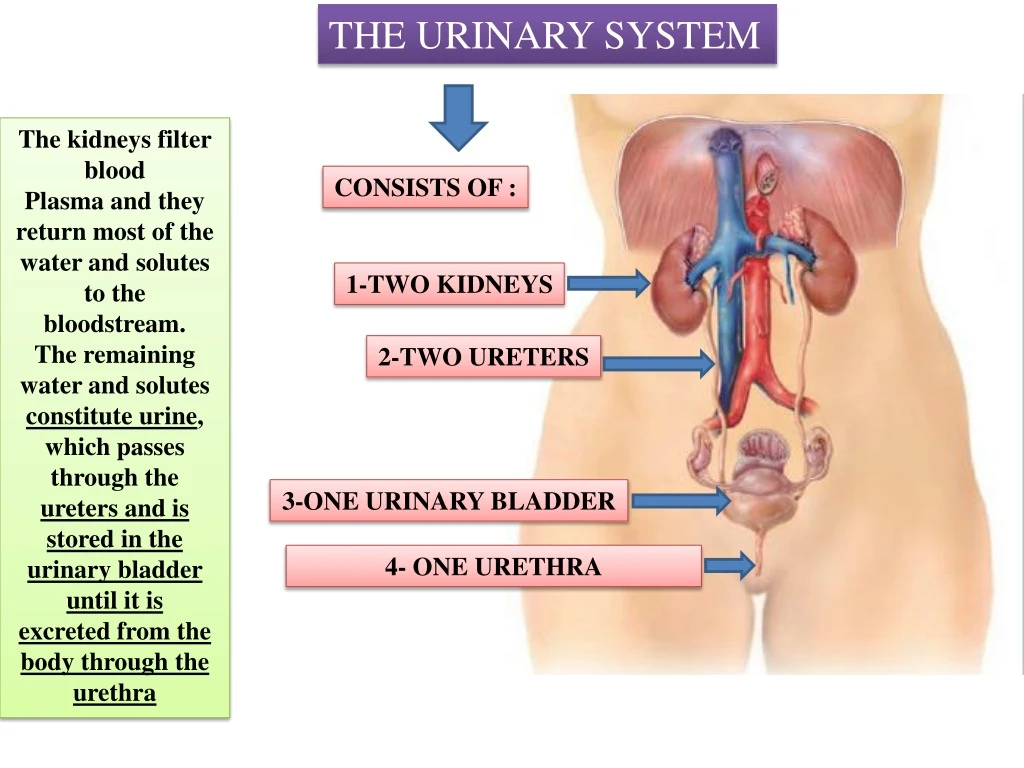
THE URINARY SYSTEM The kidneys filter blood Plasma and they return most of the water and solutes to the bloodstream. The remaining water and solutes constitute urine, which passes through the ureters and is stored in the urinary bladder until it is excreted from the body through the urethra CONSISTS OF : 1-TWO KIDNEYS 2-TWO URETERS 3-ONE URINARY BLADDER 4- ONE URETHRA
External Anatomy of the Kidneys A typical adult kidney is: 10–12 cm long 5–7 cm wide It has a concave medial border Near the center of the concave border is THE RENAL HILUM through which the ureter emerges from the kidney along with blood vessels, lymphatic vessels, and nerves Three layers of tissue surround each kidney Thedeep layer, the renal capsule The middle layer, the adipose capsule The superficial layer, the renal fascia
Internal Anatomy of the Kidneys A frontal section through the kidney reveals two distinct regions: 1-a superficial, called the renal cortex 2-deep, inner region called the renal medulla The renal medulla consists of several cone-shaped renal pyramids. Each pyramid has a base (wider end) faces the renal cortex, and an apex (narrower end), called a renal papilla, points toward the renal hilum The renal cortex extends from the renal capsule to the bases of the renal pyramids and into the spaces between them. Those portions of the renal cortex that extend between renal pyramids are called renal columns • Together, the renal cortex and renal pyramids of the renal medulla constitute • THE PARENCHYMA • Within the parenchyma are the • functional units of the kidney—about 1 million microscopic structures called NEPHRONS
The Nephron Parts of a Nephron Nephrons are the functional units of the kidneys. consists of two parts: 1-a renal corpuscle where blood plasma is filtered, And 2- a renal tubule into which the filtered fluid passes. The two components of a renal corpuscle are the a-glomerulus(capillary network) and the b-glomerular(Bowman’s) Capsule a double-walled epithelial cup that surrounds the glomerular capillaries. Blood plasma is filtered in the glomerular capsule, and then the filtered fluid passes into the renal tubule,
THE RENAL TUBULE CONSISTS OF 1) proximal convoluted tubule 2) loop of Henle (nephron loop) 3) distal convoluted tubule The distal convoluted tubules of several nephrons empty into a single collecting duct. Collecting ducts then unite and converge into several hundred large papillary ducts papillary ducts drain into the minor calyces and major calyces Each kidney has 8 to 18 minor calyces and 2 or 3 major calyces. From the major calyces, urine drains into a single large cavity called the renal pelvis and then out through the ureter to the urinary bladder
Locations of the urinary bladder URINE TRANSPORTATION, STORAGE, AND ELIMINATION Ureters Each of the two ureters transports urine from the renal pelvis of one kidney to the urinary bladder. The ureters are 25–30 cm long Urinary Bladder The urinary bladder is a hollow, muscular organ situated in the pelvic cavity posterior to the pubic symphysis. Urinary bladder capacity averages 700–800 mL in females, it is anterior to the vagina and inferior to the uterus In males, it is directly anterior to the rectum;
Urethra The urethra is a small tube leading from the internal urethral orifice in the floor of the urinary bladder to the exterior of the body. In females, it has a length of 4 cm The opening of the urethra to the exterior, the external urethral orifice, is located between the clitoris and the vaginal opening Very Short …easily can be infected In males, the urethra also extends from the internal urethral orifice to the exterior, but its length and passage through the body are considerably different than in females The male urethra first passes through the prostate, then through the deep muscles of the perineum, and finally through the penis, a distance of about 20 cm
A Foley catheter is a flexible tube that is often passed through the urethra and into the bladder.


