Download
1 / 96
1.47k likes | 3.8k Vues
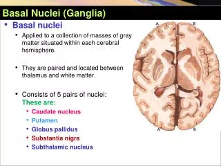
Basal Nuclei (Ganglia). Basal nuclei A pplied to a collection of masses of gray matter situated within each cerebral hemisphere. They are paired and located between thalamus and white matter . Consists of 5 pairs of nuclei: These are: Caudate nucleus Putamen Globus pallidus

E N D
Basal Nuclei (Ganglia) • Basal nuclei • Applied to a collection of masses of gray matter situated within each cerebral hemisphere. • Theyarepairedandlocatedbetweenthalamusandwhitematter. • Consists of 5 pairs of nuclei: These are: • Caudate nucleus • Putamen • Globuspallidus • Substantianigra • Subthalamic nucleus
Basal Nuclei (Ganglia) • Basal nuclei • Consists of 5 pairs of nuclei: These are: • Caudate nucleus • Putamen • Globuspallidus • Substantianigra • Subthalamic nucleus
Basal Nuclei (Ganglia) • Consists of 5 pairs of nuclei: These are: • Caudate nucleus • Putamen • Globuspallidus • Substantianigra • Subthalamic nucleus
Archistriatum Basal Nuclei (Ganglia) Amygdaloid body
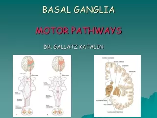
Function of BasalNuclei Basically the activity of basal nuclei begins by information received from sensory cortex, thalamus, substantianigra, and red nucleus, according to thoughts of mind. • These information is integrated within corpus striatum and channeled within globuspallidus and outflow back to motor areas of cerebral cortex, and other motor areas in brain stem. • Thus the basal nuclei can control muscular movement through its effect on cerebral cortex. • So basal nuclei assist in regulation of voluntary movement and learning of motor skills.
Functions of BasalNuclei • 1- Design of plans, which convert thoughtsand ideas into motor actions: to produce a coordinated organized purposeful movement. e.g. dressing. • Determining the timing and scale ofmovement: to what extent the movement will be fast, and how long it will last. • Storage of motor programs of familiar motor actions: e.g. signature.
Functions of NucleiBasales (GangliaBasale) • The basal ganglia are associated with a variety of functions, including : • Voluntary motor control • Procedural learning relating to routine behaviors or "habits" such as bruxism(gnashing of teeth), eye movements, and cognitive, emotional functions. • Action selection, that is, the decision of which of several possible behaviors to execute at a given time. • Basal ganglia exert an inhibitory influence on a number of motor systems, and that a release of this inhibition permits a motor system to become active.
CaudateNucleus • Large C-shaped or comma-shaped mass of grey matter. • Lies in close relation to lateral ventricle. • It has a Head, Body, and Tail.
CaudateNucleus • Large C-shaped or comma-shaped mass of grey matter. • Lies in close relation to lateral ventricle. • It has a Head, Body, and Tail.
CaudateNucleus • Head :( Anterior) Large, & rounded and forms the lateral wall of anterior horn of lateral ventricle. • It is continuous inferiorly with putamen of lentiform Nucleus. • Body : Long &narrow continuous with head at the interventricular foramen. • It forms part of the floor of body of lateral ventricle. • Tail : Long & narrow, and lies in the roof of inferior horn of lateral ventricle. • It is connected anteriorly with Amygdaloid nucleus.
Connections of CorpusStriatum • Afferent Fibers ( Input): • I- Corticostriate Fibers: From all parts of cerebral cortex (mostly from sensory- motor cortex) axons pass to caudate nucleus and putamen. • Glutamate is the neurotransmitter of this fibers. • II-Thalamostriate Fibers : From intralaminar nuclei of thalamus axons pass to caudate nucleus and putamen. • III- Nigrostriate Fibers : Axons from Substantianigra of midbrain pass to caudate nucleus and putamen. • Neurotransmitter is Dopamine. • IV_Brain stem StrialFibers : Ascending fibers from brain stem end in caudate nucleus & putamen. • Serotonin is the neurotransmitter. • It is believed that the last 2 groups are inhibitory in function.
EfferentFibers (Output) • Striatopallidal fibers: • These fibers pass from corpus striatum (caudate nucleus & putamen) to globuspallidus. • Gamma-aminobutyric acid (GABA) is the neurotransmitter. • Striatonigral fibers: • These fibers pass from caudate nucleus & putamen to Substantianigra. • Some fibers use GABA as a neurotransmitter, and others use substance p.
LentiformNucleus • It is a mass of grey matter wedge-shaped or (biconvex lens). • It has 2 capsules, external capsule laterally & internal capsule medially. • Internal capsule separates between lentiform nucleus laterally & caudate nucleus and thalamus medially. • External capsule separates between lentiform nucleus and Claustrum.
LentiformNucleus • It is divided into putamen & globuspallidus. • Putamen:Larger darker lateral portion. • Globuspallidus : Smaller, lighter medial portion. • Inferiorly putamen is continuous with the head of caudate nucleus.
LentiformNucleus • It is divided into putamen & globuspallidus. • Putamen:Larger darker lateral portion. • Globuspallidus : Smaller, lighter medial portion. • Inferiorly putamen is continuous with the head of caudate nucleus.
Basal Nuclei (Ganglia) • Substantianigra • Located in midbrain between cerebral crus and tegmentum. • Looks dark owing to melanin pigment containing neurons.. • Consists of two parts: • Pars compacta • Pars reticularis
SubstantiaNigra Aquaductus mesencephali • Tr. corticospinalis • Tr. corticonuclearis Tectum mesencephalicum Pars posterior (tegmentum mesencephalicum) Pedunculus cerebri Pc Substantia nigra Crus cerebri (pars anterior) Pr
Cross section obtained from the level of superior colliculus Nucleus tractus mesencephalici nervi trigeminalis Strata (grisea et alba) colliculi superioris Nucleus oculomotorius accessorius Lemniscus medialis Substantia nigra, Pars compacta Nucleus ruber Fibrae parietotemporopontinae Fibrae corticonucleares Substantia nigra, Pars reticularis Fibrae corticospinales Fibrae frontopontinae Fila radicularia nervi oculomotorii Substantia Nigra
Substantia Nigra • Afferents: • Mostcomefromneostriatum. • Most of theafferentfibersterminate in SNPR, but someend in SNPCas well.
Substantia Nigra • Efferents: • Pars compacta contains dopaminergic neurons. • Efferents arising from SNPCterminate in neostriatum.
Substantia Nigra • Efferents: • Pars reticularis’containsGABAergicneurons. • Efferents arising from SNPRterminate in: • : • Thalamus • Superiorcolliculus • Tegmentalnucleus • Pedunculopontinenucleus
Basal Nuclei (Ganglia) • Subthalamic nucleus • The most lower nucleus of the subthalamus. • Extents to transition zone between the tegmentum of the midbrain and subthalamus. • Located dorsolateral upper end of the SN, medial to the internal capsule, and in upper lateral part of the thalamus.
Subthalamic Nucleus • Afferents • GPE • Centromediannucleus • Parafascicularnucleus • Tegmentalnucleus • Pedunculopontinenucleus
Subthalamic Nucleus • Afferents • GPE • Centromedian nucleus • Parafascicular nucleus • Tegmental nucleus • Pedunculopontine nucleus
Subthalamic Nucleus • Efferents • GPe and GPi • Substantia nigra
SubthalamicNucleus • Efferents • GPeandGPi • Substantianigra
Nuclei Basales Bağlantıları Connections of BasalNuclei (Ganglia) • Importants features: • Efferents from cerebral cortex to striatum are activator (use glutamate). • Neurons in the striatum are inhibitory in nature.. • Neurons in the SNrand GPm are also inhibitory in nature. • Efferents of the thalamus stimulate the cerebral cortex.
Connections of BasalNuclei (Ganglia) • Directpathway: • Cortical cells project excitatory inputs to the striatum, which in turn projects inhibitory neurons onto the cells of the SNr-GPi complex. • The SNr-GPi complex projects directly onto the thalamus through the inhibitory ansalenticularispathway. • The striatal inhibition of the SNr-GPi complex coupled with SNr-GPi inhibition of the thalamus therefore results in a net reduction of inhibition of the thalamus via the striatum. Glutamate GABA GABA Glutamate
Connections of BasalNuclei (Ganglia) • Directpathway: • The thalamus projects excitatory glutamatergic neurons to the cortex itself. • The direct pathway, therefore, results in the excitation of the motor cortex by the thalamus. Once stimulated, the cortex projects its own excitatory outputs to the brain stem and ultimately muscle fibers via the lateral corticospinal tract. • The following diagram depicts the direct pathway: Glutamate GABA GABA Glutamate • Cortex (stimulates) → Striatum (inhibits) → "SNr-GPi" complex (less inhibition of thalamus) → Thalamus (stimulates) → Cortex (stimulates) → Muscles, etc. → (hyperkinetic state)